Figures & data
Figure 1 Ultrasound biomicroscopy images.
Notes: (A) Ultrasound biomicroscopy image depicting anterior segment structures in a normal eye. (B) Ultrasound biomicroscopy showing keratolenticular adhesion (arrow) with cataract in a case of Peter’s anomaly. The image demonstrates the utility of anterior segment imaging for pediatric cataract evaluation.
Abbreviations: C, cornea; I, Iris; ACa, anterior capsule; L, lens.
Abbreviations: C, cornea; I, Iris; ACa, anterior capsule; L, lens.
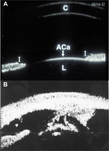
Figure 2 Examination of family members.
Notes: (A) Undilated exam that is apparently normal; (B) dilated exam of the same patient revealed punctate cortical opacities.
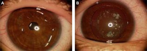
Figure 3 Total cataract with central anterior capsular plaque.
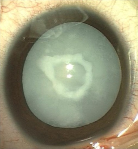
Figure 4 Anterior polar cataract.
Notes: (A) Aniridia with dot-like anterior polar cataract (arrow); (B) pyramidal cataract.
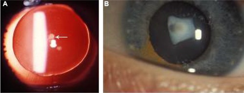
Figure 5 Oil-droplet cataract in galactosemia.
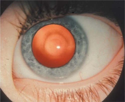
Figure 6 Subluxated lens in a child with Marfan’s syndrome.
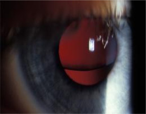
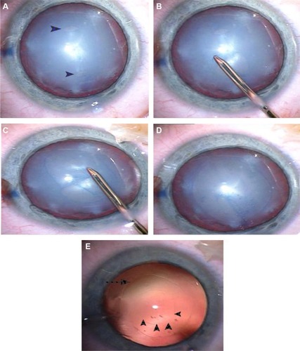
Figure 7 The technique of two-incision push–pull anterior capsulorhexis.
Notes: (A) Two stab incisions are made approximately 4.5–5.0 mm apart in the anterior capsule with a microvitreoretinal blade (arrowheads). (B) Grasping the distal flap of the proximal anterior capsule with capsulorhexis forceps and pushing toward the distal stab incision, making a semicircular rhexis; (C) similarly, the proximal flap of the distal stab incision is then grasped and pulled toward the proximal stab incision. (D) Complete continuous curvilinear capsulorhexis. (E) Anterior (arrow) and posterior two-incision push–pull rhexis (arrowheads) under retroillumination.