Figures & data
Figure 1 Photograph of the 23G RUIDO fiberscope (FiberTech Co., Ltd., Tokyo, Japan).
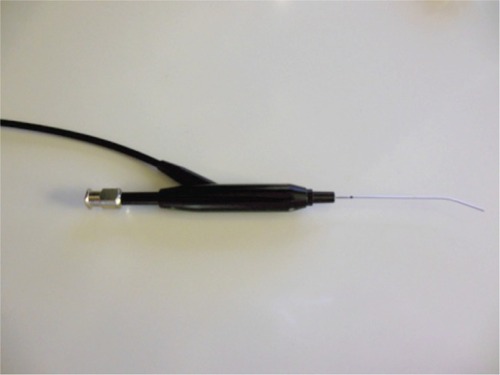
Figure 2 Dacryoendoscopic image of the lacrimal sac. The wall of the lacrimal sac is smooth with no fibrosis. There is a dimple (arrow) in the lacrimal sac.
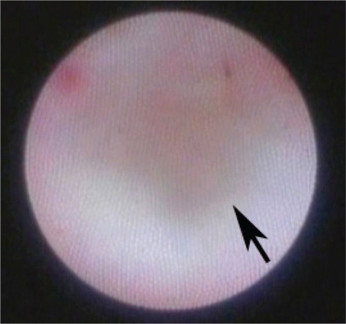
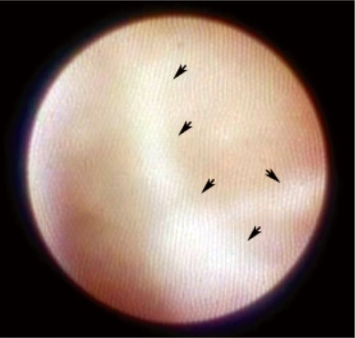
Figure 3 Dacryoendoscopic image showing fibrosis in the lacrimal sac (arrows). The lumen is to the left of the fibrosis.