Figures & data
Table 1 Comparison of characteristics and findings before and after intravitreal injection of bevacizumab in groups with branch retinal vein occlusion and diabetic retinopathy
Figure 1 Best-corrected visual acuity before and after intravitreal injection of bevacizumab.
Abbreviations: BCVA, best-corrected visual acuity; BRVOME, branch retinal vein occlusion-associated macular edema; DME, diabetic macular edema; IVB, intravitreal bevacizumab; logMAR, logarithm of the minimum angle resolution.
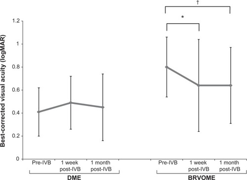
Figure 2 Foveal thickness before and after intravitreal injection of bevacizumab.
Abbreviations: BRVOME, branch retinal vein occlusion-associated macular edema; DME, diabetic macular edema; FT, foveal thickness; IVB, intravitreal bevacizumab.
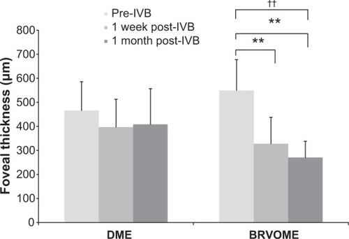
Figure 3 Mean blur rate in the retinal artery, retinal vein, optic nerve head, and choroid before and after intravitreal injection of bevacizumab.
Abbreviations: BRVOME, branch retinal vein occlusion-associated macular edema; DME, diabetic macular edema; IVB, intravitreal bevacizumab; MBR, mean blur rate; ONH, optic nerve head.
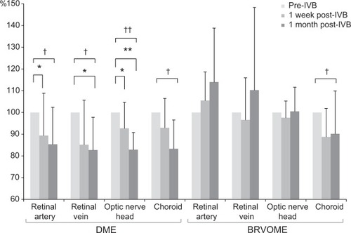
Figure 4 Relationship between foveal thickness and mean blur rate in eyes with diabetic macular edema.
Abbreviations: FT, foveal thickness; IVB, intravitreal bevacizumab; MBR, mean blur rate.
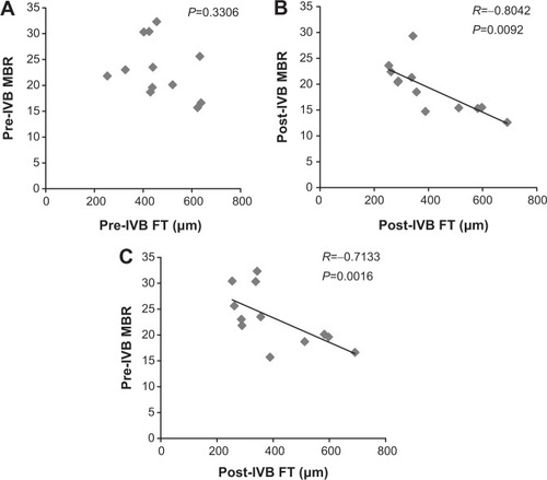
Figure 5 Relationship between foveal thickness and mean blur rate in eyes with branch retinal vein occlusion-associated macular edema.
Abbreviations: FT, foveal thickness; IVB, intravitreal bevacizumab; MBR, mean blur rate.
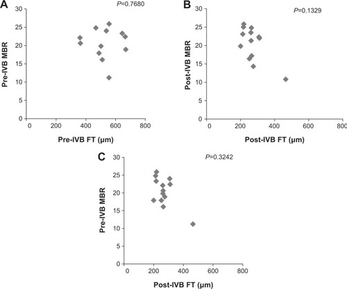
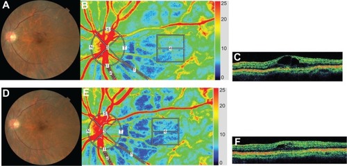
Figure 6 Representative eye with diabetic macular edema.
Abbreviations: DME, diabetic macular edema; FT, foveal thickness; IVB, intravitreal bevacizumab; LSFG, laser speckle flowgraphy; MBR, mean blur rate; OCT, optical coherence tomography.
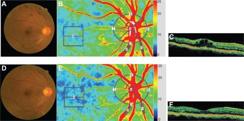
Figure 7 Representative eye with branch retinal vein occlusion-associated macular edema.
Abbreviations: BRVOME, branch retinal vein occlusion-associated macular edema; FT, foveal thickness; IVB, intravitreal bevacizumab; LSFG, laser speckle flowgraphy; MBR, mean blur rate; OCT, optical coherence tomography.