Figures & data
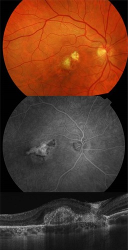
Figure 1 Juxtafoveolar subretinal choroidal neovascularization with subretinal fluid adjacent to the choroidal rupture 2 months after vitrectomy demonstrated by optical coherence tomography and fluorescein angiography.
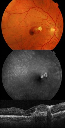
Figure 2 Recurrence of the choroidal neovascularization activity with a central macular thickness of 349 μm.
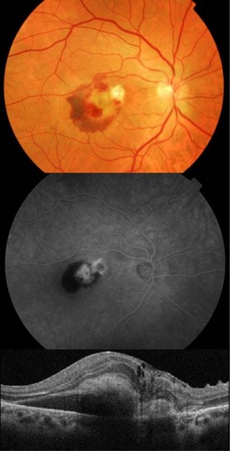
Figure 3 Spectral-domain optical coherence tomography confirmed reduction of the choroidal neovascularization activity with a central macular thickness of 275 μm and disappearance of the retinal hemorrhage. Leakage from the choroidal neovascularization in the fluorescein angiography had ceased.