Figures & data
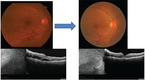
Figure 1 Case one: fundus and optical coherence tomographic images of a 72-year-old woman with a CRVO before and after treatment of systemic hypertension with a calcium blocker.
Notes: At the first visit her BP was 169/96 mmHg with no medications, and her fundus showed an impending CRVO with macular edema. Her VA was 20/50. One month after the treatment of systemic hypertension, her macular edema completely disappeared, and her VA was improved to 20/20 with no recurrence for at least 1 year.
Abbreviations: BP, blood pressure; CRVO, central retinal vein occlusion; VA, visual acuity.
Abbreviations: BP, blood pressure; CRVO, central retinal vein occlusion; VA, visual acuity.
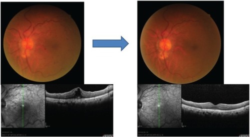
Figure 2 Case two: fundus and optical coherence tomographic images of a 62-year-old woman with BRVO before and after treatment of her systemic hypertension with an angiotensin II blocker.
Notes: Her BP was 165/97 mmHg at the first visit. Macular edema decreased, and her VA improved from 20/40 to 20/20 after treatment.
Abbreviations: BP, blood pressure; BRVO, branch retinal vein occlusion; VA, visual acuity.
Abbreviations: BP, blood pressure; BRVO, branch retinal vein occlusion; VA, visual acuity.
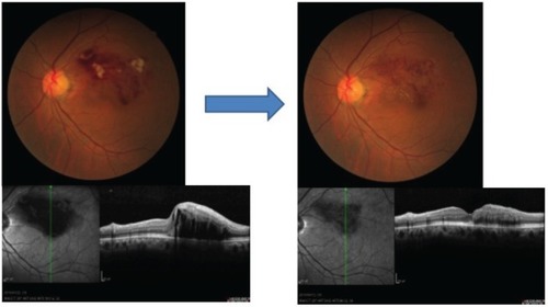
Figure 3 Case three: fundus and optical coherence tomographic images of a 71-year-old man with a BRVO before and after treatment of systemic hypertension with angiotensin receptor blocker.
Notes: His BP was 165/87 mmHg at the first visit. Macular edema was not present with greatly improved VA from 20/50 to 20/20 1 month after treatment.
Abbreviations: BP, blood pressure; BRVO, branch retinal vein occlusion; VA, visual acuity.
Abbreviations: BP, blood pressure; BRVO, branch retinal vein occlusion; VA, visual acuity.