Figures & data
Table 1 Epidemiological data and outcomes of all subjects
Figure 1 Case 4 (pre-injection). The corneal abscess caused by Aspergillus flavus has spread to most of the grafted cornea.
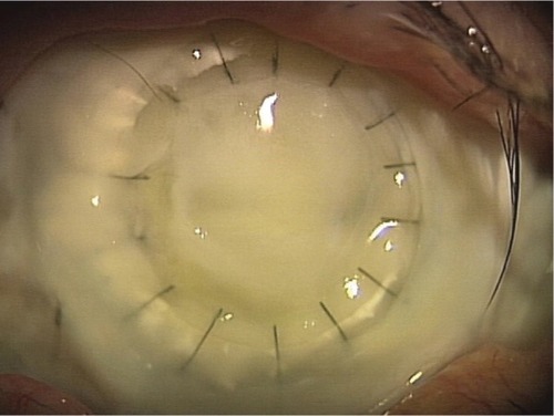
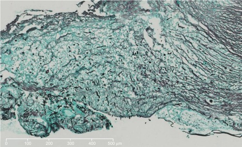
Figure 2 Case 4. Pathological findings of the removed cornea (Grocott’s staining). A substantial amount of fungi are observed just above the Descemet’s membrane.
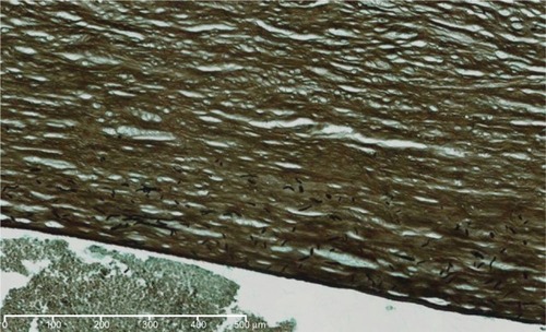
Figure 3 Optical coherence tomography images of Case 1 (pre- and post-injection).
Notes: Intrastromal fluid location just after the injection of voriconazole can be seen in the right picture. The high-density area is limited to the outermost two-thirds or three-fourths of the corneal stroma (white arrows).
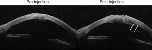
Figure 4 Case 5 (the anterior picture at the initial visit).
Note: The hyphate ulcer and the corneal perforation are observed.
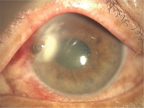
Figure 5 Case 5 (pathological findings of the removed cornea) (Grocott’s staining).
Note: Hyphae is observed in the deep layers of the corneal stroma.