Figures & data
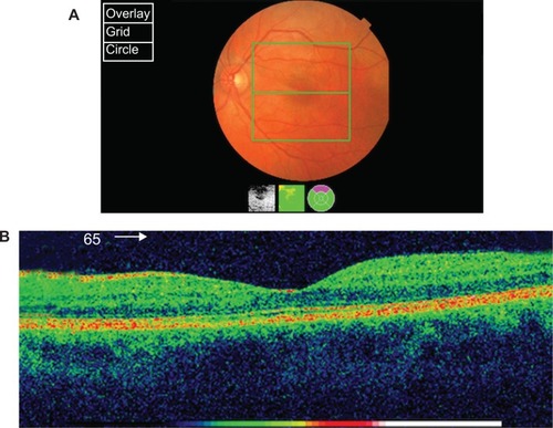
Figure 1 Fundoscopy and OCT at presentation.
Notes: (A) Fundoscopy at presentation showing superotemporal commotio retinae and an abnormal cream foveal discoloration. (B) The OCT of left macula at presentation shows outer photoreceptor segment disruption, RPE inter-digitation with some outer and inner segment foveal disruption, and intra-retinal edema at the outer nuclear layer.
Abbreviations: OCT, optical coherence tomography.
Abbreviations: OCT, optical coherence tomography.
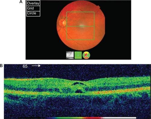
Figure 2 Fundoscopy and OCT after 3 months.
Notes: (A) Fundoscopy after 3 months suggests almost complete resolution of the retinal layers. (B) OCT findings after 3 months suggest almost complete resolution of the retinal layers edema with a small discontinuity in the inner and outer segments adjacent to the fovea.
Abbreviations: OCT, optical coherence tomography.
Abbreviations: OCT, optical coherence tomography.