Figures & data
Figure 1 Ultrawide-field fundus photography of gyrate atrophy (Optos Imaging System). Extensive chorioretinal atrophy with remaining central macula islands at the posterior poles demarcated by pigmented borders.
Notes: (A) Right eye; (B) left eye. Optos® (Dunfermline, United Kingdom).
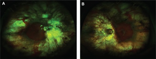
Figure 2 (A) Grayscale visual field testing of the left eye; (B) grayscale visual field testing of the right eye (Carl Zeiss Meditec AG, Jena, Germany).
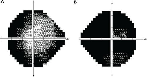
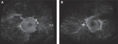
Figure 3 Ultrawide-field fluorescein angiography of gyrate atrophy (Optos Imaging System).
Notes: (A) Right eye; (B) left eye. Optos® (Dunfermline, United Kingdom).