Figures & data
Table 1 Direct photocoagulation to treat macular edema associated with BRVO: patient demographics
Figure 1 Reduction in the central foveal thickness (CFT) after direct photocoagulation.
Notes: (A) The line graph shows the changes in the CFT 3 months and 6 months after direct photocoagulation. *P<0.01 by one-way analysis of variance and the Tukey’s test. (B) The bar graph shows a comparison of the baseline and final CFTs. *P<0.01, by the paired t-test.
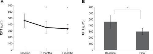
Table 2 Visual outcomes after direct photocoagulation for chronic macular edema associated with BRVO
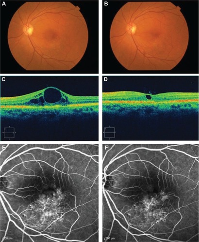
Figure 2 Images of the left eye of a 68-year-old woman with branch retinal vein occlusion (BRVO).
Notes: A fundus photograph (top), optical coherence tomography (OCT) image (middle), and fluorescein angiography (FA) image (bottom) were obtained at baseline (A, C, E) and 3 months (B, D, F) after direct photocoagulation. (D) OCT shows decreased macular edema after direct photocoagulation compared to the baseline (C). FA shows decreased microaneurysms and leakage after direct photocoagulation. The best-corrected visual acuity (BCVA) improved from 40/200 to 60/200.