Figures & data
Figure 1 There was white perivascular hard exudate (yellow arrow) along the vessels in both eyes, and multiple patches of cotton-wool spots around the disk and macula in her left eye.
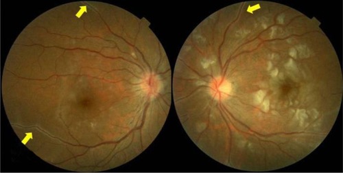
Figure 2 (A) After pulse therapy, there were increasing cotton-wool spots and multiple arterioles narrowing at the macula in her right eye and confluent macular cotton-wool spots presenting like cherry-red spots with attenuated smaller arterioles in her left eye. (B) At the early phase in the macula, a small branch of a capillary nonperfusion zone was observed in the right eye, and multiple branches of arterioles were occluded in the left eye. (C) At the late phase in the macula, perivascular leakage of multiple arterioles was observed in both eyes. There was an extensive capillary nonperfusion zone in the left macula.
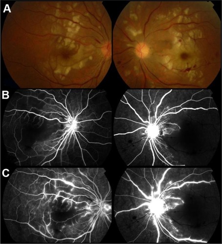
Figure 3 The relationship of visual acuity and steroid use under sub-tenon injection and intravenous way.
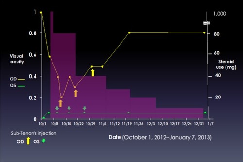
Figure 4 (A) After sub-Tenon’s injection of triamcinolone acetonide 50 mg/week in both eyes for 3 weeks, there were obviously decreasing soft exudates in the macula in both eyes. (B) At the late phase in the macula, there was less perivascular leakage in each eye and a smaller capillary nonperfusion area in the right eye.
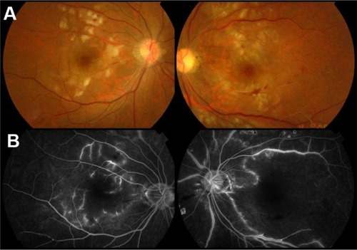
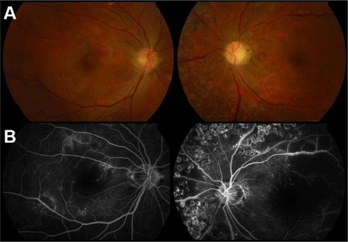
Figure 5 (A) There were no cotton-wool spots over posterior pole in either eye. The left disk seemed mildly waxy pale. (B) At the late phase in the macula, there was a smaller nonperfusion area in the right eye.