
Figures & data
Figure 1 Anterior segment photography of the right eye.
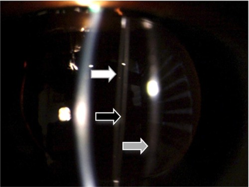
Figure 2 Comparative ultrabiomicroscopies of both eyes. (A) Right eye. The intraocular lens is displacing the iris forward. There is an echo-negative space between the intraocular lens and the distended posterior capsule. (B) Left eye. A normally placed intraocular lens. There is no abnormal contact with any surrounding structure.
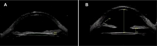
Figure 3 Anterior segment photography of the right eye after Nd:YAG capsulotomy.
Abbreviation: Nd:YAG, neodymium-doped yttrium aluminum garnet.
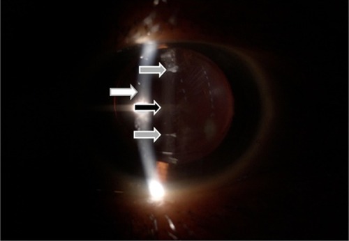
Figure 4 Anterior segment photography of the right eye after Nd:YAG capsulotomy.
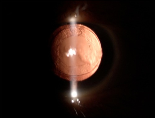
Figure 5 Ultrabiomicroscopy of the right eye after Nd:YAG capsulotomy. The capsular block syndrome is resolved. There is no contact between the intraocular lens and the iris.
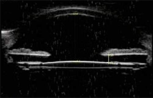
Figure 6 Anterior segment photography of the right eye using a Scheimpflug-based camera.
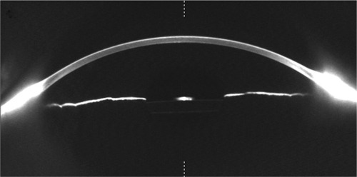
Figure 7 Anterior segment photography of the left eye using a Scheimpflug-based camera.
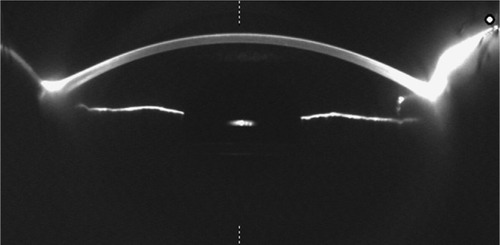
Table 1 Classification according to time of the development of capsular block syndrome
Table 2 Pathophysiological classification of CBS as proposed by Kim and Shin