Figures & data
Figure 1 Findings in a 48-year-old Japanese man with oculocutaneous albinism. His best-corrected visual acuity was 2/100 with −14.0 diopters in the right eye.
Notes: (A) External photograph of the right eye showing white cilia and a depigmented iris. (B, C) Fundus photographs at initial examination showing diffuse depigmentation. The right eye had a foveal hemorrhage (B). (D, E) Fluorescein angiographic images at the initial visit showing relative hypofluorescent retinal and choroidal vessels because of the lack of melanin pigment in the retinal pigment epithelium. The right eye has an area of hypofluorescence corresponding to the foveal hemorrhage (D). (F) Indocyanine green angiographic image of the right eye at the initial visit does not show any findings suggestive of choroidal neovascularization. (G) Optical coherence tomographic image corresponds to the arrow in Figure (D). A spectral-domain optical coherence tomographic image of the right eye at the initial visit shows an absence of the foveal pit, and the presence of a subretinal hyperreflective lesion corresponding to the foveal hemorrhage.
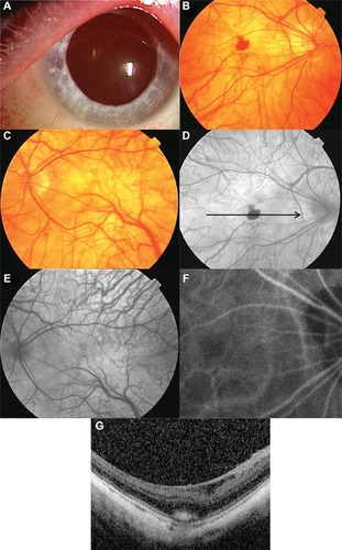
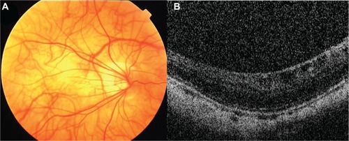
Figure 2 Three months after onset. The patient’s best-corrected visual acuity has not improved.
Notes: (A) Fundus photograph of the right eye showing that the foveal hemorrhage has regressed. (B) Spectral-domain optical coherence tomographic image of the right eye showing an absence of a subretinal hyperreflective lesion corresponding to the foveal hemorrhage.