Figures & data
Figure 1 Slit-lamp photograph of the case.
Notes: (A) Preoperative slit-lamp photograph showing an opaque and edematous cornea due to endothelial dysfunction. (B) One year postoperatively, the corneal graft clarity was excellent, with a high endothelial cell density (4,032 cells/mm2, 6.0% decrease from preoperative donor cell measurements).
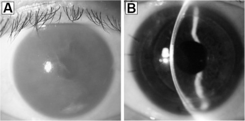
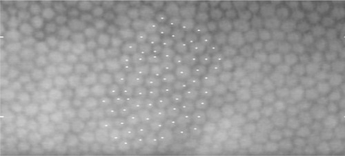
Figure 2 Preoperative analysis of the infant donor endothelial cells.
Notes: The endothelial cell density of the donor tissue was as high as 4,291 cells/mm2, with no striae.