Figures & data
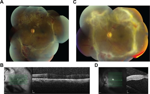
Figure 1 Progression of diabetic tractional retinal detachment, by fundus photography and optical coherence tomography (OCT) of the left eye after a unilateral, right eye intraoperative intravitreal bevacizumab injection.
Notes: Fundus photograph (A) and OCT (B) of the left eye before right eye intraoperative intravitreal bevacizumab injection, showing vitreoretinal adhesions and attached macula. Fundus photograph (C) and OCT (D) of the left eye after right eye intraoperative intravitreal bevacizumab injection, showing decreased perfusion of the neovascular tissue and progression of tractional retinal detachment.