Figures & data
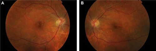
Figure 1 Retinography 12 hours post partum.
Notes: Bilateral optic disc edema, narrowing, and irregularity of retinal arteries with arteriovenous nicking, diffuse cotton wool spots, intraretinal and subretinal transudates, and multiple superficial and deep retinal hemorrhages. (A) right eye; (B) left eye.
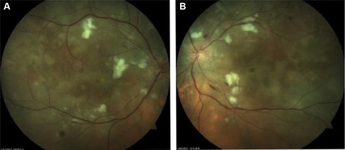
Figure 2 Fluorescein angiogram in early and late phases 12 hours post partum.
Notes: Fluorescein angiogram revealed delayed perfusion of the choriocapillaris in both eyes. The posterior pole and peripheral retina presented multiple capillary dilations, focal venous bleeding, capillary nonperfusion areas, retinal hemorrhages, and late leakage in the optic disc. Multiple areas of focal choroidal ischemia (Elschnig’s spots). (A) and (B) right eye; (C) and (D) left eye.
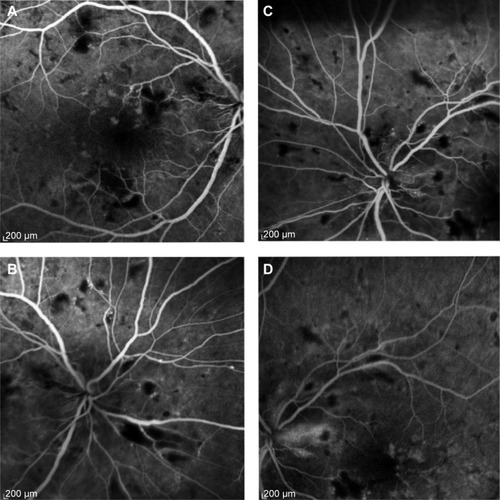
Figure 3 Optical coherence tomography 12 hours post partum.
Notes: Retinal edema, retina serous detachments presenting with a hyperreflective posterior surface of the detached retina, subretinal hyperrreflective lesions corresponding to subretinal transudates, and a thickened peripapillary retinal nerve fiber layer corresponding to optic disc edema. (A) and (B) right eye; (C) and (D) left eye. Insets are infrared fundus image with corresponding OCT.
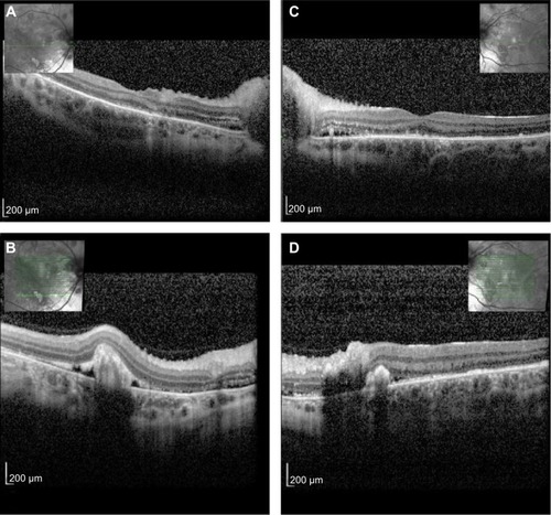
Figure 4 Follow-up examination at 6 months after the acute phase.
Notes: Complete resolution of previous serous detachments, diffuse arteriolar constriction, and multiple yellowish subretinal discoloration spots. (A) right eye; (B) left eye.