
Figures & data
Figure 1 Fundus imaging.
Notes: (A) Color fundus photograph (Topcon TRC 50EX), (B) red free imaging (Heidelberg Spectralis), (C) infrared imaging (Heidelberg Spectralis), and (D) fundus autofluorescence (Heidelberg Spectralis).
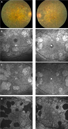
Figure 2 Spectral domain optical coherence tomography images of both eyes through the retinal crystals.
Notes: The yellow arrows demonstrate the location of the crystals in the retina in a region of preserved outer retina (upper panel) and atrophic outer retina (lower panel).
