Figures & data
Figure 1 Slit-lamp photo before and after DMEK.
Notes: (A) Before surgery, severe bullous keratopathy secondary to argon laser iridotomy was noted in the right eye. (B) One week after DMEK, a crystal clear cornea was observed. The patient’s best corrected visual acuity improved rapidly to 25/20.
Abbreviation: DMEK, Descemet membrane endothelial keratoplasty.
Abbreviation: DMEK, Descemet membrane endothelial keratoplasty.
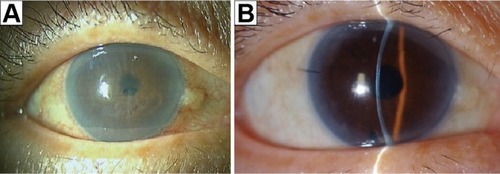
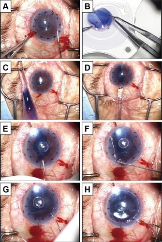
Figure 2 Surgical technique of endoillumination-assisted DMEK.
Notes: (A) After removal of the host epithelial membrane for better visualization of the anterior chamber, the host DM was removed under viscoelastic materials that filled the anterior chamber. (B) The donor graft was prepared using the submerged cornea with a backgrounds away technique. (C and D) The harvested and stained DM roll stained by trypan blue was then inserted into the anterior chamber using a DMEK shooter. (E–G) The DMEK roll was correctly oriented with the endothelium side facing down. A small air bubble was then used to unfold the DM graft. To obtain further visualization, oblique light via an endoillumination probe held by an assistant surgeon was used. This technique improved the contrast between the blue-stained DM roll and the background of the dark brown iris. (H) Finally, the anterior chamber was filled with air to attach the DM graft completely to the posterior stromal surface.
Abbreviations: DMEK, Descemet membrane endothelial keratoplasty; DM, De scemet membrane.
Abbreviations: DMEK, Descemet membrane endothelial keratoplasty; DM, De scemet membrane.