Figures & data
Figure 1 Serial OCT scans, right eye in left column and left eye in right column.
Notes: Outer cystic retinal edema prior to cataract extraction (A and B). Increased cystic edema OU and SRF OD after cataract extraction; choroidal thickness demarcated by white arrows (C and D). Resolved edema and SRF after starting acetazolamide (E and F). Mild return of edema at temporal disc border when carbonic anhydrase inhibitors were stopped (G and H).
Abbreviations: OCT, optical coherence tomography; OU, oculus uterque (both eyes); SRF, subretinal fluid; OD, oculus dexter (right eye).
Abbreviations: OCT, optical coherence tomography; OU, oculus uterque (both eyes); SRF, subretinal fluid; OD, oculus dexter (right eye).
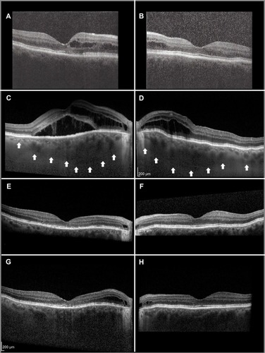
Figure 2 Composite fundus images.
Notes: Color photographs show healthy discs, temporal peripapillary crescents, and choroidal folds (A and B). Fluorescein angiograms demonstrated no macular retinal epithelial decompensation, although the crescents stained in the late phase (C and D). Late hyperfluorescence suggests hyperpermeability in the posterior pole on an ICGA (E and F).
Abbreviation: ICGA, indocyanine green angiogram.
Abbreviation: ICGA, indocyanine green angiogram.
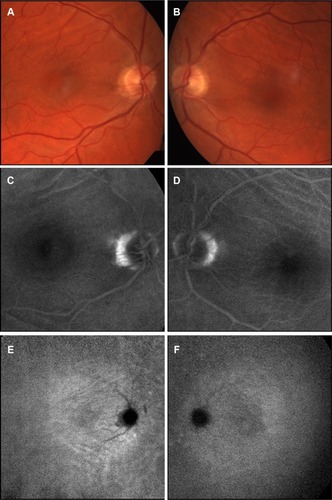
Figure 3 Beneath a cystic space in the outer peripapillary retina.
Notes: The outer neurosensory retina and retinal pigment epithelium are attenuated, as seen on this magnified view of .
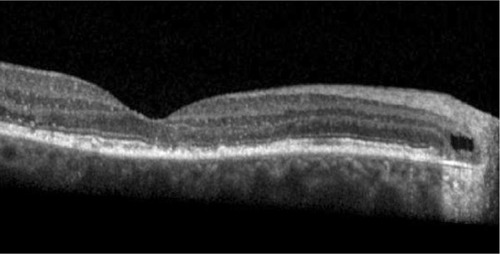
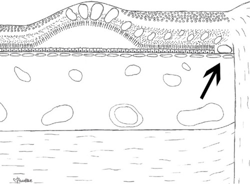
Figure 4 Artist rendition of the macula.
Notes: This artist rendition of the macula shows proposed site of fluid leakage from the choroid into the retina (arrow). Fluid passes from the choroid through Bruch’s membrane at the temporal crescent, where the fluid passes directly into the retina because of the attenuation of the RPE and photoreceptor layers. The fluid gravitates toward the macula, where it passes through the external limiting membrane into the subfoveal neurosensory space.
Abbreviation: RPE, retinal pigment epithelium.
Abbreviation: RPE, retinal pigment epithelium.