Figures & data
Figure 1 Diagram showing the location of three collimated beams of radiation passing through the inferior sclera (green circles) to avoid the crystalline lens, during stereotactic radiotherapy.
Note: Image courtesy of Oraya (Oraya Therapeutics, Newark, CA, USA).
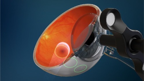
Figure 2 Illustration to show the attenuation effect of the three collimated beams to deliver 90% of the desired radiation dose to a 4 mm diameter at the macula.
Notes: Outside the 4 mm diameter zone, the radiation delivered diminishes rapidly. Image reproduced with modification, courtesy of Oraya (Oraya Therapeutics, Newark, CA, USA).
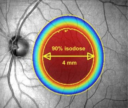
Figure 3 The room set-up of the IRay system.
Notes: The operator controls (left) are separated from the radiotherapy machine (right) and patient by a lead-lined glass screen, allowing the operator to view the patient. Image courtesy of Oraya (Oraya Therapeutics, Newark, CA, USA).
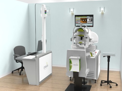
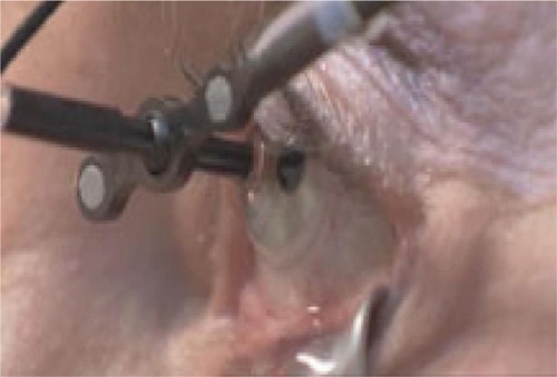
Figure 4 A contact lens, with light suction mechanism, is used to keep the eye in position during treatment.
Note: Copyright © 2011. Petrarca R, Jackson TL. Radiation therapy for neovascular age-related macular degeneration. Clin Ophthalmol. 2011;5:57–63.Citation22