
Figures & data
Figure 1 Preoperative right fundus and OCT imaging in Case 1.
Notes: Peripheral retinal detachment with subretinal strands is visible. The macula appears uninvolved, but OCT shows the absence of the inner and outer segment junction line, indicating spontaneous attachment.
Abbreviation: OCT, optical coherence tomography.
Abbreviation: OCT, optical coherence tomography.
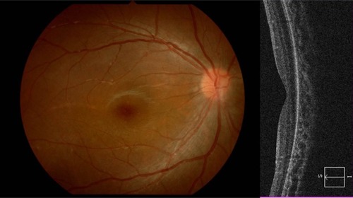
Figure 2 Preoperative left fundus and optical coherence tomography imaging in Case 2.
Note: Flat retinal detachment with subretinal strands and macular involvement are visible.
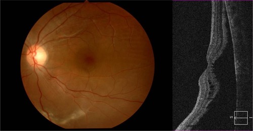
Figure 3 Image of surgical procedure.
Notes: Left: Intraoperative view of the inserted chandelier. Right: Intraoperative fundus image under chandelier illumination and the view of cryoretinopexy.
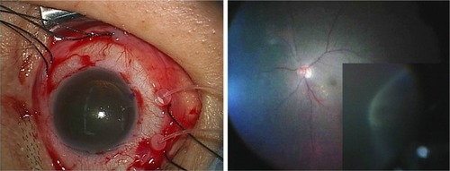
Table 1 Clinical characteristics of the three cases