Figures & data
Figure 1 Changes of BCVA after surgery.
Notes: Mean change in visual acuity up to month 12; (A) all patients and (B) ischemic and non-ischemic patients, from baseline. BCVA values at 3, 6, and 12 months after the vitrectomy were significantly better than preoperative BCVA values. Vertical bars are ±1 SEM. *P<0.05, Wilcoxon signed-rank test.
Abbreviations: BCVA, best-corrected visual acuity; logMAR, logarithm of the minimum angle of resolution; SEM, standard error of the mean.
Abbreviations: BCVA, best-corrected visual acuity; logMAR, logarithm of the minimum angle of resolution; SEM, standard error of the mean.
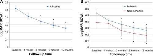
Table 1 Patient demographic data and preoperative clinical status
Figure 2 Changes in CFT after surgery.
Notes: Mean change in CFT up to month 12; (A) all patients and (B) ischemic and non-ischemic patients from baseline. Mean CFT decreased significantly 1 month after surgery, and postoperative CFT was significantly decreased from the baseline thickness at all times evaluated. Vertical bars are ±1 SEM. *P<0.05, Wilcoxon signed-rank test.
Abbreviations: CFT, central foveal thickness; SEM, standard error of the mean.
Abbreviations: CFT, central foveal thickness; SEM, standard error of the mean.
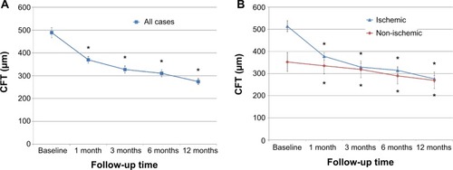
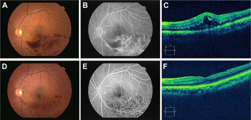
Figure 3 Clinical findings before and after surgery.
Notes: (A) Preoperative fundus photograph; (B) early venous phase fluorescein angiogram, and (C) spectral domain optical coherence tomographic image of a 62-year-old women with ischemic branch retinal vein occlusion. Best-corrected visual acuity was 6/20, and central foveal thickness was 424 μm. (D) Postoperative color fundus photograph, (E) early venous phase fluorescein angiogram, and (F) spectral domain optical coherence tomographic image showing an almost normal foveal contour 3 months after surgery. Best-corrected visual acuity was 20/20 and central foveal thickness was 239 μm.