
Figures & data
Figure 1 Visual evoked potential of a healthy participant.
Notes: Normal visual evoked potential waveform parameters on both sides. (A) Visual evoked potential of the right eye. (B) Visual evoked potential of the left eye. (C) Parameters of both eyes.
Abbreviations: Ampl, amplitude; Div, division; Fz, lead placed at the mid line frontal; Lat, latency; Oz, lead placed at the mid line occipital; rec, recording; Stim, stimulating.
Abbreviations: Ampl, amplitude; Div, division; Fz, lead placed at the mid line frontal; Lat, latency; Oz, lead placed at the mid line occipital; rec, recording; Stim, stimulating.
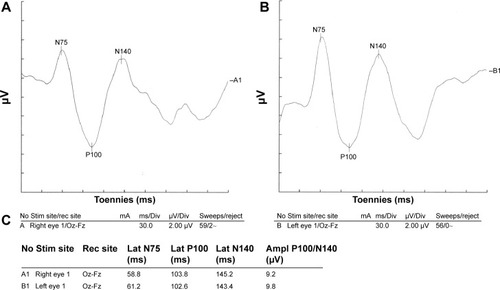
Figure 2 Visual evoked potential of a patient with COPD.
Notes: Left side P100 latency was prolonged; Conclusion: normal right side visual evoked potential waveform parameters and mild demyelinating type lesion on left side optic pathway. (A) Visual evoked potential of the right eye. (B) Visual evoked potential of the left eye. (C) Parameters of both eyes.
Abbreviations: Ampl, amplitude; COPD, chronic obstructive pulmonary disease; Div, division; Fz, lead placed at the mid line frontal; Lat, latency; Oz, lead placed at the mid line occipital; rec, recording; Stim, stimulating.
Abbreviations: Ampl, amplitude; COPD, chronic obstructive pulmonary disease; Div, division; Fz, lead placed at the mid line frontal; Lat, latency; Oz, lead placed at the mid line occipital; rec, recording; Stim, stimulating.
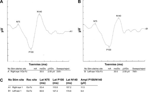
Table 1 Results of VEP studies in two groups