Figures & data
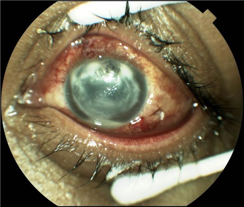
Figure 1 Acute-onset postoperative endophthalmitis (note the sutured corneal wound and hypopyon).
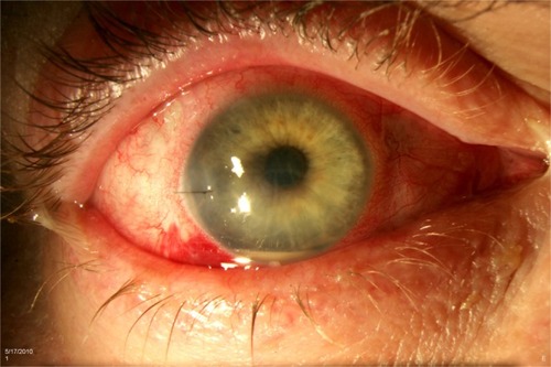
Figure 2 Delayed-onset (chronic) postoperative endophthalmitis (note the small hypopyon and peripheral intracapsular infiltrates).
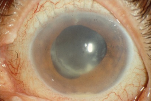
Figure 3 Bleb-related endophthalmitis (note the purulent filtering bleb and hypopyon).
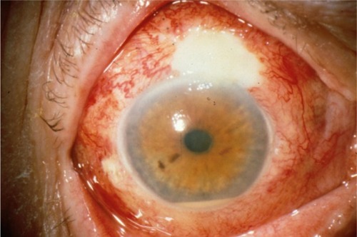
Figure 4 Endophthalmitis following intravitreal injection (note the chemosis and hypopyon).
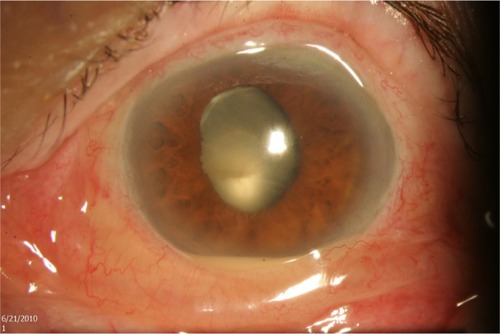
Figure 5 Posttraumatic endophthalmitis (note the sutured corneal wound and hypopyon).