
Figures & data
Figure 1 Nine measurement points of choroidal thickness and the area between the 3-papilla diameter and 9-papilla diameter circles (midperipheral area) in which abnormal autofluorescence abnormalities were investigated.
Notes: The nine measurement points of choroidal thickness are indicated by stars. The short double-headed arrow indicates 3-papilla diameter and the long one indicates 9-papilla diameter.
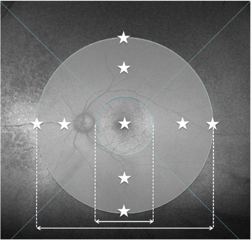
Table 1 Patient demographics
Figure 2 Comparison of choroidal thickness.
Notes: (A) The mean choroidal thickness was higher in CVH(+) AMD eyes than in CVH(-) AMD eyes at the fovea and the superior, inferior, and nasal points. (B) Choroidal thickening was greater in eyes with PCV than in eyes with tAMD at the fovea and inferior point. Asterisks indicate significant differences (P<0.05, t-test). Bars indicate SD.
Abbreviations: CVH(−), choroidal vascular hyperpermeability-negative; CVH(+), choroidal vascular hyperpermeability-positive; tAMD, typical age-related macular degeneration; PCV, polypoidal choroidal vasculopathy; AMD, age-related macular degeneration; SD, standard deviation.
Abbreviations: CVH(−), choroidal vascular hyperpermeability-negative; CVH(+), choroidal vascular hyperpermeability-positive; tAMD, typical age-related macular degeneration; PCV, polypoidal choroidal vasculopathy; AMD, age-related macular degeneration; SD, standard deviation.
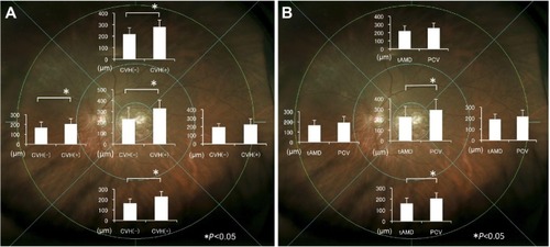
Figure 3 Two representative cases of CVH(+) AMD showing FAF abnormalities in the midperipheral fundus.
Notes: (A) Arrows show hyperfluorescence and arrowheads show hypofluorescence in the temporal midperipheral fundus. (B) Arrows show hyperfluorescence in the temporal midperipheral fundus.
Abbreviations: CVH(+), choroidal vascular hyperpermeability-positive; AMD, age-related macular degeneration; FAF, fundus autofluorescence.
Abbreviations: CVH(+), choroidal vascular hyperpermeability-positive; AMD, age-related macular degeneration; FAF, fundus autofluorescence.

Table S1 Previous treatments of patients