Figures & data
Figure 1 Case 1: a 44-year-old male.
Notes: (A) Nine days after the appearance of the initial symptoms. The cornea of the left eye had a large area of circular epithelial defects with an irregular surface. (B) The epithelial defect widened with an overhanging edge on the tenth day. (C) The cornea of the right eye started to present with multiple full-layer epithelial defects, smaller than the ones in the left eye. (D) The previous defect of the cornea re-epithelialized, and subepithelial corneal infiltrates were observed.
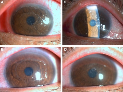
Figure 2 Case 2: A 15-year-old male.
Notes: (A) Full-layer cornea epithelial detachment with a scrolling over-hanging edge appeared in the left eye of a 15-year-old male. (B) The epithelial defect started to heal rapidly during the next 2 days.
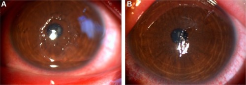
Figure 3 Case 3: a 5-year-old female.
Notes: (A) Severe papillary congestion with patchy subconjunctival hemorrhage in the conjunctiva and an epithelial defect on the cornea in the right eye of a 5-year-old female. (B) The large epithelial defect started to heal after 3 days.
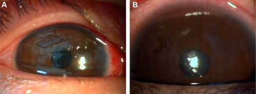
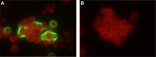
Figure 4 Detection of adenovirus in the HEp-2 cells derived from swab samples of patients by indirect immunofluorescence assay. The cytoplasm and nuclei were stained red by Evans blue, and green fluorescent signals were on adenovirus-infected cells.
Notes: (A) Adenovirus-infected cells were detected with anti-adenovirus antibody. (B) Uninfected cells were the negative control.