
Figures & data
Figure 1 Showing the distribution of Chrysomya bezziana worldwide.
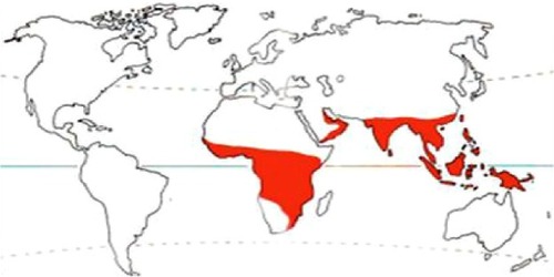
Figure 2 Lesion at the medial canthus, with free-moving maggots.
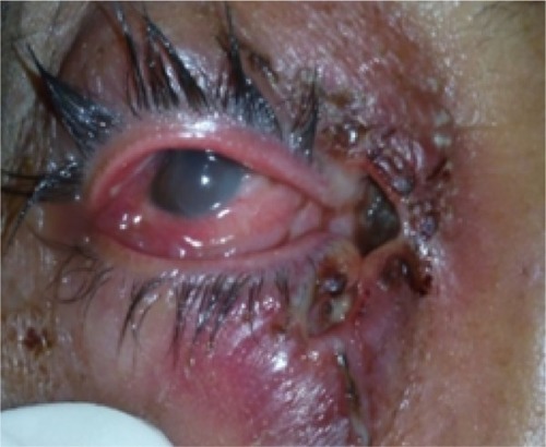
Figure 3 CT scan orbit showing no signs of bone involvement, with normal sinuses.
Abbreviation: CT, computed tomography.
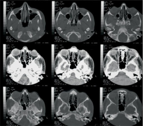
Figure 4 Mechanical removal of maggots with wound debridement.
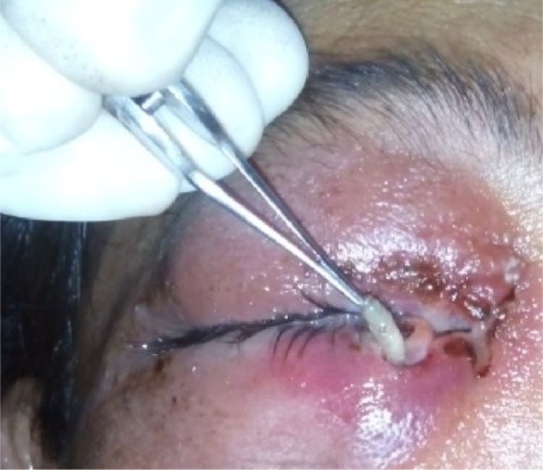
Figure 5 Follow up at 2 weeks, with completely healed wound.
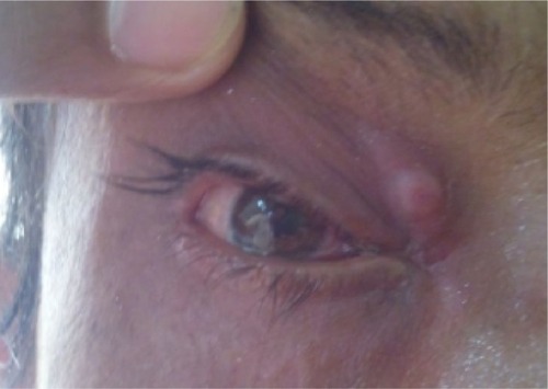
Figure 6 Maggots removed from patient’s wound.
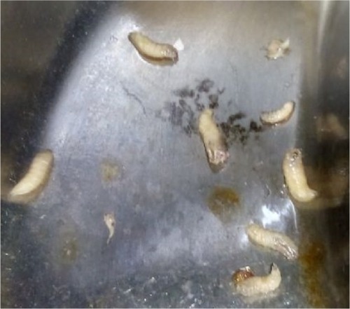
Figure 7 Mouth hooks with cuticular spines around body of larvae.
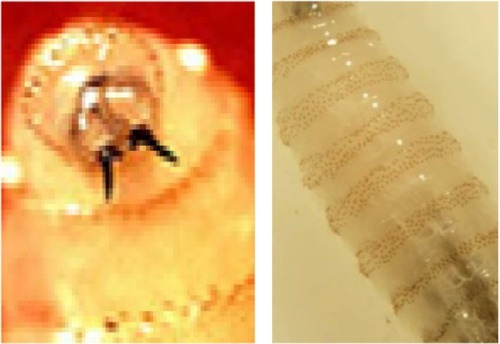
Figure 8 Incomplete posterior spiracular peritreme of Chrysomya bezziana.
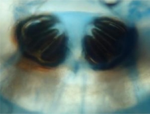
Table 1 Agents used in treatment of myiasis