Figures & data
Table 1 Key inflammatory mediators driving BRB breakdown in diabetic macular edema
Table 2 Comparative efficacy and safety of dexamethasone and fluocinolone acetonide intravitreal implants in diabetic macular edema
Table 3 Summary of Phase II and Phase III clinical trials of dexamethasone intravitreal implant in diabetic macular edema
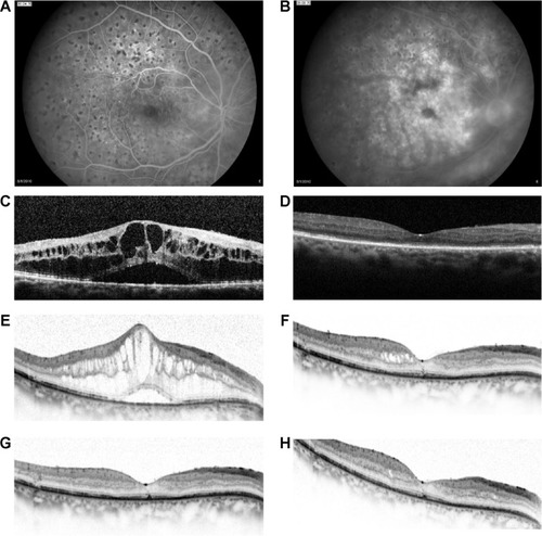
Figure 1 A 64-year-old male diagnosed with diabetic macular edema in the right eye.
Abbreviation: BCVA, best-corrected visual acuity.
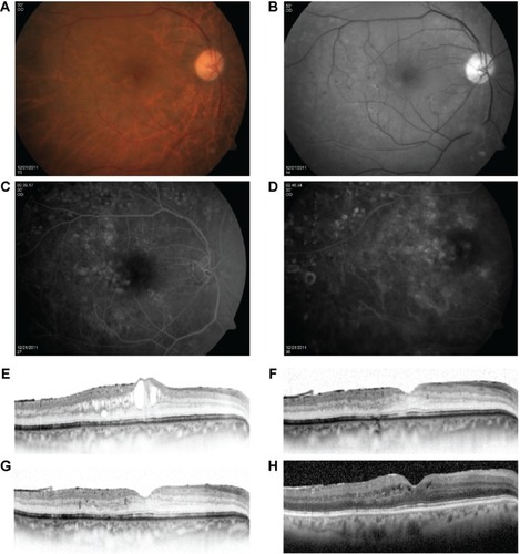
Figure 2 A 62-year-old patient with cystoid macular edema in the left eye previously treated with three monthly injections of intravitreal bevacizumab.
Abbreviation: BCVA, best-corrected visual acuity.
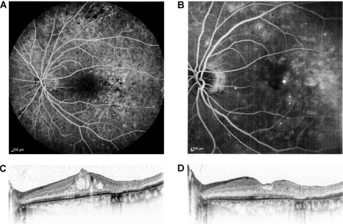
Table 4 Most frequently reported adverse events in Phase III studies of dexamethasone intravitreal implant in the treatment of diabetic macular edema
Figure 3 A 72-year-old patient with diabetic macular edema in the right eye previously treated with three monthly injections of intravitreal bevacizumab.
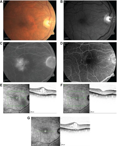
Figure 4 A 65-year-old female with regressed proliferative diabetic retinopathy in the right eye previously treated with three monthly injections of intravitreal bevacizumab.
Abbreviation: BCVA, best-corrected visual acuity.