Figures & data
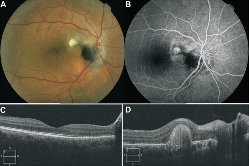
Figure 1 Fundus color image, fluorescein angiography and optical coherence tomography findings in patient’s right eye at initial examination.
Notes: (A) Color image of the right eye fundus at first consultation, showing a deep black melanocytoma (arrow) occupying the inferotemporal half of the optic disk extending temporally to the adjacent retina (note the striated margins) and apparently to the inferior juxtapapillary choroid; subretinal choroidal neovascular complex (arrowhead) near the superotemporal margin of the optic disk, with multiple fine subretinal folds radiating from it, and two retinal folds crossing horizontally through the central macula; a myriad of fine reflective subretinal lipid deposits under the macula and nasal half of the posterior pole extending beyond the temporal vascular arcades and densely concentrated subretinal lipids at the inferior margin of the optic disk; and moderate optic disk edema and dilatation and tortuosity of superior temporal and nasal retinal veins. (B) Early arteriovenous phase fluorescein angiogram showing evident epipapillar and peripapillar retinal capillary telangiectasia, extending four-disk diameters superotemporally at both sides of the superotemporal retinal vein (arrows). (C) Venous phase fluorescein angiogram of the same eye, showing leakage from the choroidal neovascular complex (arrows) and from telangiectatic retinal capillaries of the superior juxtapapillar area (arrowheads). (D) Time domain optical coherence tomography scan passing through the fovea and choroidal neovascular complex in the same eye (arrowheads), showing shallow neurosensory detachment (arrows) and intraretinal edema in the superonasal quadrant of the macula.
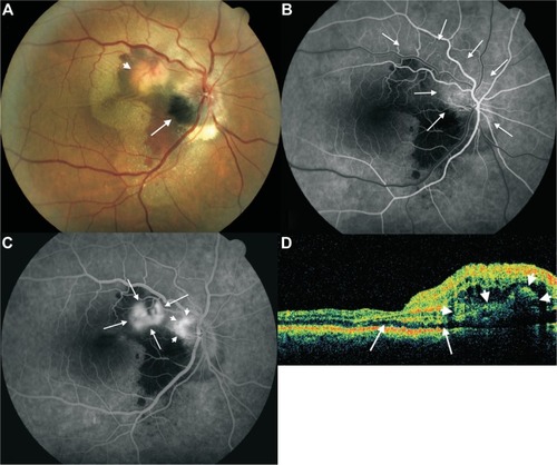
Figure 2 Post treatment fundus color image, fluorescein angiography and optical coherence tomography findings.
Notes: (A) Color image of the right eye fundus of the same patient 3 years after treatment showing complete resolution of subretinal fluid, lipid deposits and hemorrhage, and a fibrotic scar at the level of the choroidal neovascular complex; an apparent subretinal extension from the superotemporal margin of the tumor extends as a tongue toward the choroidal neovascular scar; inframacular and infrapapillar subretinal punctuate pigment dispersion is also observed, and adjacent choroidal component melanocytoma is now visible at the inferior margin of the optic disk. (B) Venous phase of fluorescein angiogram showing impregnation of choroidal neovascular scar and no capillary telangiectasia and juxtapapillar and optic disk leakage. (C) Spectral domain optical coherence tomography scan passing through the macula showing complete resolution of subretinal fluid and macular edema. (D) Spectral domain optical coherence tomography scan passing through choroidal neovascular scar.