Figures & data
Figure 1 Pre- and postoperative photographs.
Notes: (A and B) BGI surgery images of the patient’s right eye (OD) (A) and left eye (OS) (B) show bilateral aniridia and corneal opacification OD. The corneal diameters enlarged to 14.0×13.5 mm OD and 13.5×13.0 mm OS. (C and D) OD (C) and OS (D) images at the completion of the BGI surgeries. The BGI tubes were left longer in the anterior chamber than usual in preparation for tube retraction. (E and F) Fundus photographs OD (E) and OS (F) 14 months after BGI surgeries show obvious optic disc improvements. Disc shapes were oval, and the cup to disc ratio was 0.3 OD and 0.4 OS. (G and H) OD photographs just before the tube repositioning show the tube touching the cornea. (I–L) OD images during the tube repositioning surgery. (I and J) After making scleral flap at the inferior temporal area (green arrows), the end of the tube was pulled out through the corneal wound. (K) 10-0 Prolene straight needle was bent, used to penetrate through the tube end, and pulled under the scleral flap using the leading needle. (L) The tube was extended to the site of scleral flap by pulling the 10-0 Prolene. (M and N) OD photographs after tube repositioning with scleral fixation (M) and 3 months (N) after surgery. It should be noted that the tube end is extended to the inferior temporal direction by 10-0 Prolene and there is a space between the tube and the cornea. (O) OS photograph 18 months after BGI surgery shows the BGI tube has come slightly closer to but has not yet touched the cornea. (P and Q) Anterior segment optical coherence tomography images OD (P) and OS (Q) show that spaces exist between the cornea and the tube.
Abbreviation: BGI, Baerveldt glaucoma implant.
Abbreviation: BGI, Baerveldt glaucoma implant.
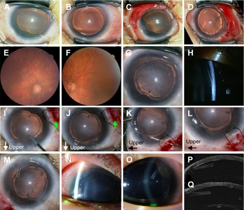
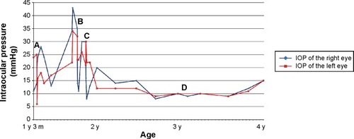
Figure 2 The intraocular pressure (IOP) time course.
Notes: All IOP measurements were performed under sedation using a Perkins applanation tonometer (Clement-Clarke, Haag-Streit UK, Essex, UK). The patient underwent the first inferotemporal trabeculotomy OS (left eye) at 15 months (A). His IOP OS decreased to around the mid-teens once, but increased to over 30 mmHg and his IOP OD (right eye) gradually increased to over 40 mmHg. Therefore, a second inferonasal trabeculotomy OS and BGI OD were performed at 21 months (B). After 1 month, BGI OS was performed (C), because the second trabeculotomy OS was not sufficiently effective. The BGI tube repositioning with scleral fixation OS was performed (D).
Abbreviations: BGI, Baerveldt glaucoma implant; y, year(s); m, month(s).
Abbreviations: BGI, Baerveldt glaucoma implant; y, year(s); m, month(s).