Figures & data
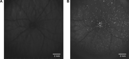
Figure 1 The DARC in vivo retinal image.
Notes: White spots represent single undergoing apoptosing RGCs. (A) Baseline image. (B) Chemically induced apoptosis captured by wide-field lens 2 hours after intravitreal administration of fluorescent-labeled annexin V. Images used an Heidelberg Retinal Angiograph Spectralis (Heidelberg Engineering, Heidelberg, Germany).
Abbreviations: DARC, detection of apoptosing retinal cell; RGCs, retinal ganglion cells.
Abbreviations: DARC, detection of apoptosing retinal cell; RGCs, retinal ganglion cells.