
Figures & data
Figure 1 Representative images of various corneal damages due to HSV1 infection.
Notes: (A) Large herpetic epithelial dendrite at graft–host junction. (B) Large subepithelial bulla due to HSV endotheliitis. (C) Ring-lipid deposit surrounding a focal HSV disciform keratitis. (D) Large geographic herpetic ulcer in HIV patient. (E) Herpetic keratouveitis with anterior chamber inflammation (layered hypopyon due to WBC accumulation), small keratic precipitates (WBC aggregates on the corneal endothelial surface), and corneal edema (due to endothelial dysfunction). (F) Large herpetic epithelial dendrite. (G) Postherpetic neurotrophic epithelial defect due to corneal nerve damage by HSV1. (H) Large herpetic corneal scar with iris incarceration to the side of corneal perforation.
Abbreviations: HSV, herpes simplex virus; WBC, white blood cell.
Abbreviations: HSV, herpes simplex virus; WBC, white blood cell.
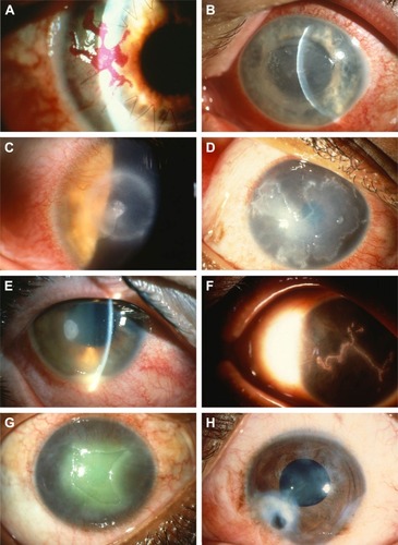
Table 1 Summary of current treatment for different subtypes