
Figures & data
Figure 1 Axial image illustrating a fully opacified (white arrow) and a fully aerated (black arrow) lacrimal sac.
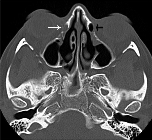
Figure 2 Axial image illustrating an opacified (small arrow) and a partially aerated (large arrow) nasal lacrimal duct.
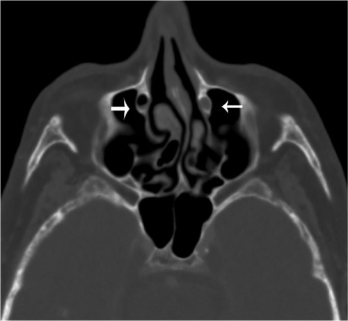
Figure 3 Coronal image illustrating an opacified (small arrow) and a fully aerated (large arrow) lacrimal sac.
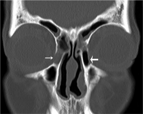
Figure 4 Coronal image illustrating an opacified nasolacrimal duct (arrow). Notes: Due to patient rotation, the contralateral duct cannot be viewed in this frame.
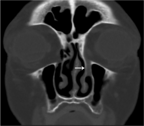
Figure 5 Coronal image illustrating a fully or partially aerated nasolacrimal duct (arrow).
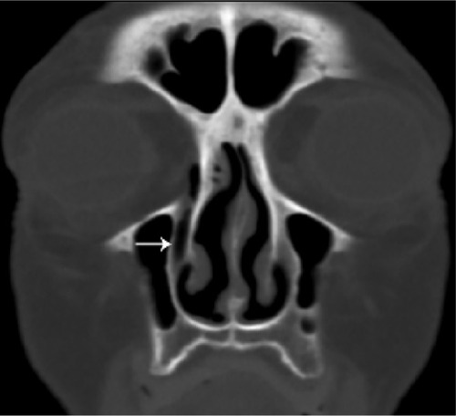
Table 1 Overall identification of air in the nasolacrimal drainage system for all patient positions (supine/upright) and scan orientations (axial/coronal), for three reviewers
Table 2 Comparison in the aeration patterns for two scan positions (upright and supine), independent of scan orientation
Table 3 The effects of positioning in a subset of four individuals who underwent both supine and upright imaging
Table 4 Nasolacrimal system aeration patterns when images are categorized by scan position (supine vs upright), location (NLS vs NLD), and scan orientation (coronal vs axial)