Figures & data
Figure 1 External photograph discloses a palpable prominence in the medial canthal area with surrounding erythema.
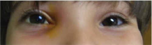
Figure 2 Magnetic resonance imaging (MRI) of patient demonstrating large ethmoidal mucocele on the right.
Notes: (A) Coronal T1 MRI, (B) sagittal T1 MRI, (C) axial T1 MRI.
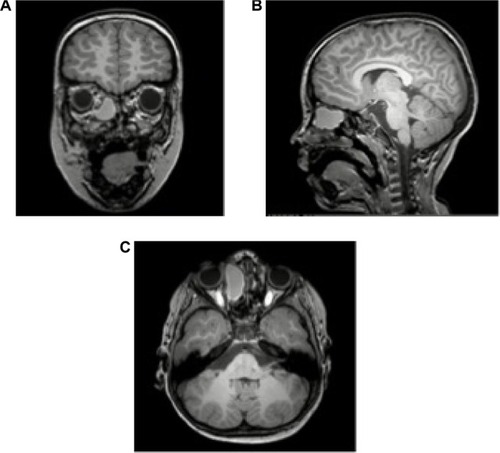
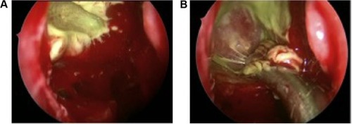
Figure 3 Images during FESS.
Notes: (A) View of sinus mucocele during FESS, (B) marsupialization of mucocele during FESS.
Abbreviation: FESS, functional endoscopic sinus surgery.
Abbreviation: FESS, functional endoscopic sinus surgery.