Figures & data
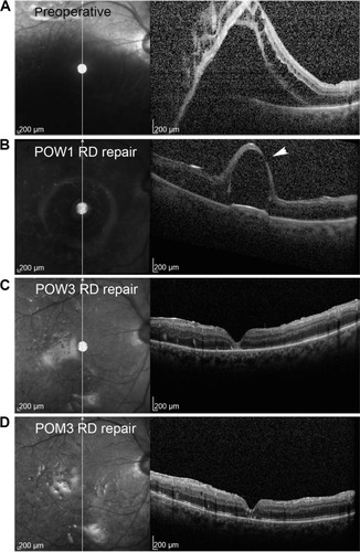
Figure 1 Spectral domain optical coherence tomographic images with corresponding scanning laser ophthalmoscopic images of the left macula.
Notes: (A) Preoperatively, the retina is detached under silicone oil. (B) At 1 week following recurrent retinal detachment repair, the retina remains attached with a large subfoveal PFCL droplet. There is a focal area of severe thinning with a possible retinal defect (arrowhead) overlying the droplet. (C) At 3 weeks following repair, without any surgical intervention, the retina remains attached, but the PFCL droplet is no longer identified and is replaced by granular changes at the outer retina with a restored foveal contour. (D) By 3 months following repair, these granular changes have improved, with visualization of outer retinal structures.
Abbreviations: PFCL, perfluorocarbon liquid; RD, retinal detachment; POW, postoperative week; POM, postoperative month.
Abbreviations: PFCL, perfluorocarbon liquid; RD, retinal detachment; POW, postoperative week; POM, postoperative month.