Figures & data
Figure 1 Color fundus photograph of the left eye demonstrates dilated, tortuous retinal veins with intraretinal hemorrhages in all four quadrants.
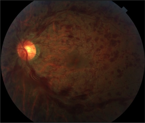
Figure 2 Fluorescein angiogram, left eye.
Notes: (A) Arterial phase of fluorescein angiography of the left eye demonstrates normal arterial filling with blockage of choroidal fluorescence by retinal hemorrhages. (B) Delayed venous filling and venous tortuosity and dilation are evident in the arteriovenous phase. (C) Normal choroidal perfusion persists through the venous phase without evidence of leakage or vasculitis.
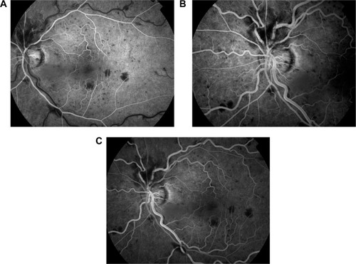
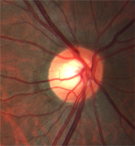
Figure 3 In the left eye, the bifurcation of the central retinal artery sits atop a segment of the central retinal vein at the site of obstruction (bracket).
Notes: The tight anatomical relationship is accentuated by an undilated nasal retinal vein firmly wrapped around the arterial bifurcation (arrowhead). The veins that emanate from underneath the artery are dilated and tortuous.
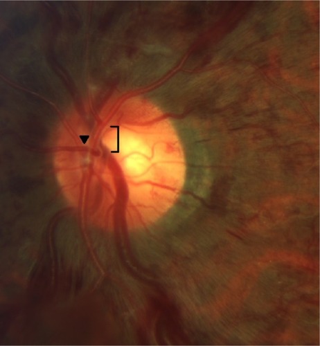
Figure 4 In the unaffected right eye, no intertwining of the vessels is seen.