Figures & data
Figure 1 Photographs showing (A) temporary globe collapse under open-cannula conditions and (B) reinflation of the eye after non-valved cannulas are occupied while working under air.
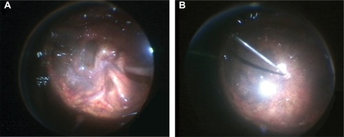
Figure 2 Experimental setup of the fluid-filled plastic eye model.
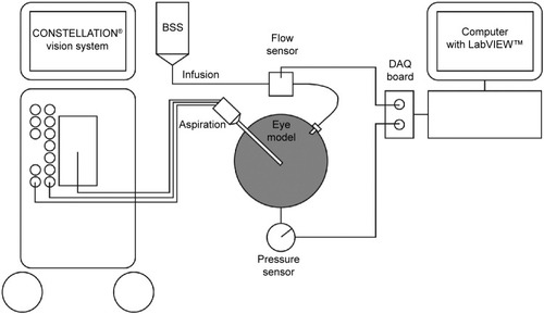
Figure 3 Intraoperative view of a valved (left) and a non-valved (right) cannula in a rabbit cadaver eye.
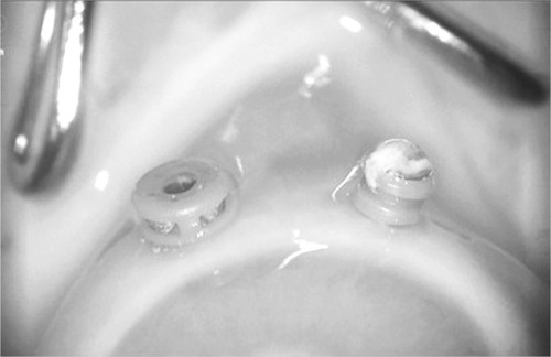
Figure 4 Experimental setup for evaluating vitreous incarceration in rabbit and porcine cadaver eyes.
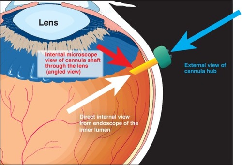
Figure 5 Intraoperative image of a rabbit cadaver eye.
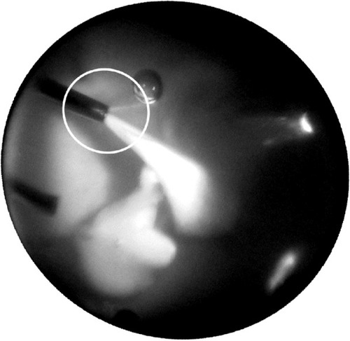
Table 1 Intraocular pressure (mmHg) in the air-filled model
Figure 6 Fluid-filled plastic eye model: change in IOP, taking the 25-gauge cannula as an example.
Abbreviation: IOP, intraocular pressure.
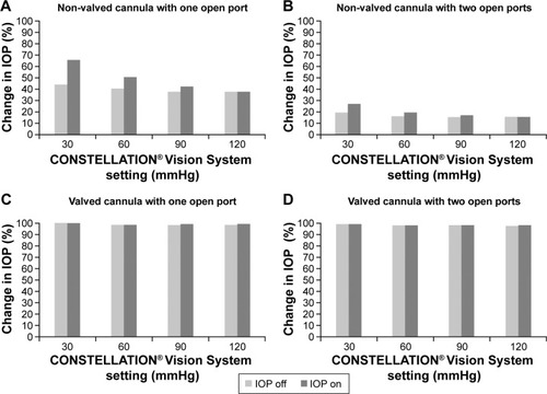
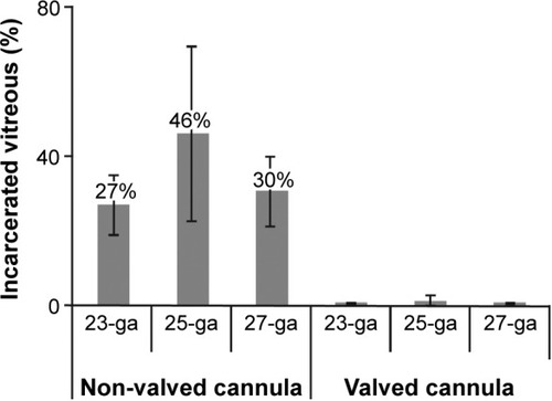
Figure 7 Vitreous incarceration in rabbit cadaver eyes.
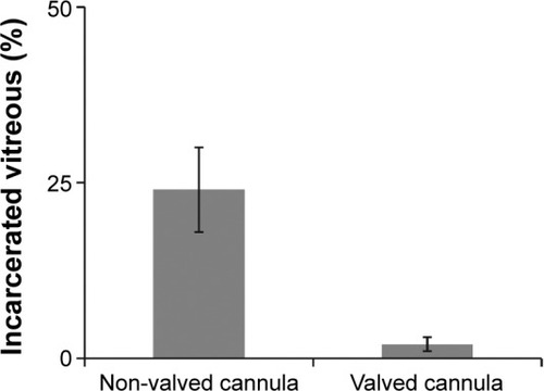
Figure 8 Vitreous incarceration in porcine cadaver eyes.