Figures & data
Figure 1 Fundus pictures of the left eye of an 11-year-old child who presented with blurred vision.
Notes: (A) At presentation, multiple subretinal yellow–white lesions were seen superiorly and at the superotemporal periphery and treated with antitubercular drugs and oral steroids (tapered over 2 months). (B) 1 month after stopping oral steroids, recurrent crops of multiple yellow–white subretinal lesions appeared at macula, which were treated with laser photocoagulation to the inferior retina and high-dose oral albendazole therapy. (C) Complete resolution of lesions was observed at 2 months. The child remains recurrence free 2 years after treatment.
Abbreviations: 20/20, Snellen visual acuity equivalent to 20/20; CFCF, visual acuity counting fingers close to face; 20/40, Snellen visual acuity equivalent to 20/40.
Abbreviations: 20/20, Snellen visual acuity equivalent to 20/20; CFCF, visual acuity counting fingers close to face; 20/40, Snellen visual acuity equivalent to 20/40.
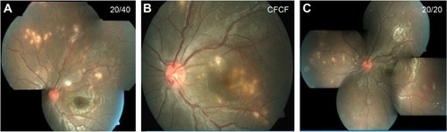
Table 1 Clinical features of patients presenting with presumed diffuse unilateral subacute neuroretinitis (DUSN)
Table 2 Age distribution of patients with DUSN
Table 3 Visual acuity at presentation
Table 4 Posterior segment findings in patients with presumed DUSN
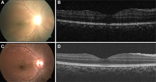
Figure 2 Fundus pictures and OCT images of early stage and chronic stage of the disease.
Notes: Fundus picture (A) and OCT (B) of right eye of a patient in the early stage of the disease showing hazy view due to vitritis, blurred disc margin, loss of foveal reflex, and macular thickening (central macular thickness =315 µm). Fundus picture (C) and OCT (D) of the same eye in the chronic stage of the disease showing resolved vitreous haze, disc pallor, and decreased macular thickening (central macular thickness =240 µm). OCT machine used was Cirrus 4000 SW version: 5.2.0.210, Carl Zeiss Meditec, Jena, Germany.
Abbreviation: OCT, optical coherence tomography.
Abbreviation: OCT, optical coherence tomography.