Figures & data
Table 1 Types of primary tumors and patients’ demographic data
Table 2 Clinical and imaging characteristics of orbital metastatic lesions
Figure 1 Imaging patterns of orbital metastasis.
Notes: (A) Magnetic resonance image showing right infiltrative metastatic lesion from breast carcinoma (arrow). (B) Computed tomography (CT) scan with contrast showing well-defined right superior orbital metastatic mass from thyroid carcinoma (arrow). (C) CT scan with contrast showing right isolated medial rectus thickening in a case of orbital metastasis from cutaneous malignant melanoma (arrow).
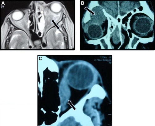
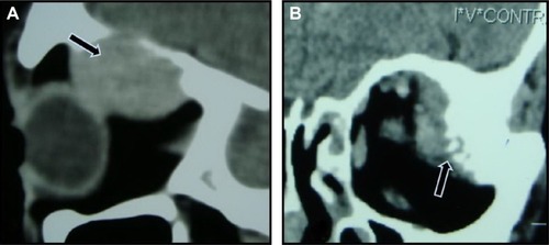
Figure 2 Computed tomography scans with contrast showing bone changes in orbital metastasis.
Notes: (A) Osteoclastic metastatic lesion from hepatocellular carcinoma (arrow). (B) Osteoblastic metastatic lesion from prostatic carcinoma (arrow).