Figures & data
Figure 1 Radial FAF at the posterior pole.
Notes: Cases 1, 3, 4, and 7 show marked radial patterns in both eyes, while the radial FAF of cases 2 and 5 exhibits low contrast in the right eye only. The radial FAF extends only to the superior temporal area in the right eye of case 6. The asterisks indicate the images without radial FAF. The images within the yellow box represent those of the study participant with a radial pattern limited to the periphery.
Abbreviation: FAF, fundus autofluorescence.
Abbreviation: FAF, fundus autofluorescence.
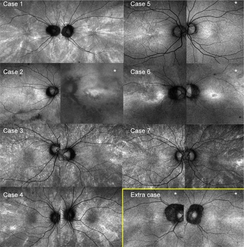
Figure 2 Peripheral radial FAF.
Notes: Except in case 5, the radial FAF is depicted better in the periphery than at the posterior pole. The images within the yellow box represent those of the study participant with a radial pattern limited to the periphery.
Abbreviation: FAF, fundus autofluorescence.
Abbreviation: FAF, fundus autofluorescence.
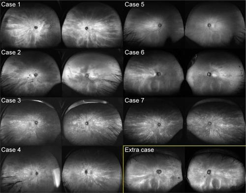
Figure 3 Visual fields of patients with radial FAF.
Notes: All eyes show visual field defects to various extents. The V4/e isopter defect of the eye in case 1 shows marked wedge-shaped constriction. Cases 1, 2, 3, and 4 were assessed with Goldmann perimetry. Case 6 was assessed using the Humphrey Field Analyzer (30-2 protocol). Visual field tests were not performed for cases 5 and 7.
Abbreviation: FAF, fundus autofluorescence.
Abbreviation: FAF, fundus autofluorescence.
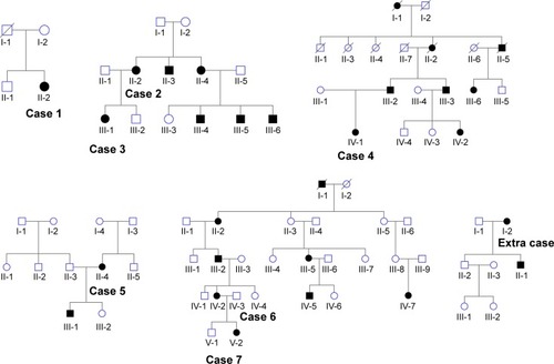
Table 1 Characteristics of the cases with radial pattern fundus autofluorescence
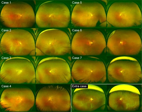
Figure 4 Color fundus photograph of patients with radial FAF.
Notes: Bone spicule pigmentation is seen only in cases 1 and 4. No eyes show the tapetal-like reflex.
Abbreviation: FAF, fundus autofluorescence.
Abbreviation: FAF, fundus autofluorescence.
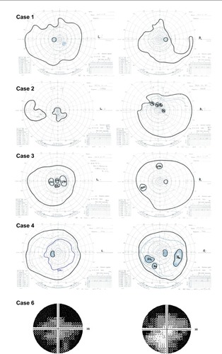
Figure 5 Family history of patients with radial FAF.
Notes: All individuals showing any symptom or fundus appearance are represented by closed marks. The extra case represents the individual with a radial pattern limited to the periphery.
Abbreviation: FAF, fundus autofluorescence.
Abbreviation: FAF, fundus autofluorescence.