Figures & data
Figure 1 The machine panel display of the Quadrant Removal Setting (Quadrant Removal Step Button enlarged) in the procedure step toolbar shows a −2 vacuum rise with linear control (angled line) of torsional amplitude.
Abbreviation: IOP, intraocular pressure.
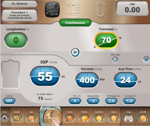
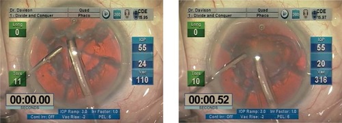
Figure 2 Video freeze frames from standard slow motion 30 frames/second video show the posterior capsule aspiration event.
Abbreviations: IOP, intraocular pressure; Vac, vacuum.
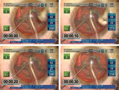
Figure 3 Video freeze frame of third quadrant removal.
Abbreviations: Vac, vacuum; IOP, intraocular pressure.
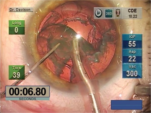
Figure 4 The machine panel display of the Epinucleus Removal Setting (Epinucleus Removal Step Button enlarged in the procedure step toolbar) shows linear control of vacuum and aspiration flow in foot position 2, which becomes fixed at their maximums in foot position 3 when linear control of continuous torsional amplitude becomes available. The iP feature is engaged but does not come into play in soft cataracts. The Epinucleus Removal Setting procedure step button is between the Sculpt and Quadrant Removal procedure step buttons in the procedure step toolbar along the bottom of the display.
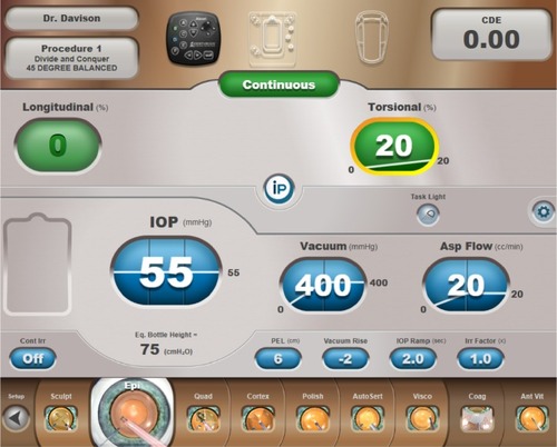
Figure 5 The machine screen display of the Sculpt Setting (Sculpt Procedure Step Button enlarged in the procedure step toolbar) shows low fixed values for vacuum and aspiration flow and linear control of continuous longitudinal tip motion.
Abbreviation: IOP, intraocular pressure.
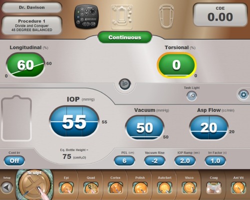
Figure 6 Soft cataract technique using the Epinucleus Removal Setting.
Abbreviations: IOP, intraocular pressure; Vac, vacuum.
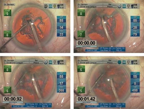
Figure 7 Removal of the second and subsequent quadrants of relatively firm soft lenses using the Quadrant Removal Setting.
Abbreviations: IOP, intraocular pressure; Vac, vacuum.