Figures & data
Figure 1 Fluorescein slit-lamp photographs of dendritic ulcers with terminal bulbs in Case 1 (A), Case 2 (B), Case 3 (C), and Case 4 (D).
Note: The area framed by the white rectangle corresponds to the confocal maps in .
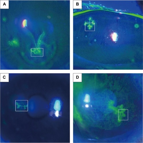
Figure 2 Subconfluent montages of in vivo confocal microscopic images at the area of dendritic lesion in Case 1 (A), Case 2 (B), Case 3 (C), and Case 4 (D). (Bar =200 μm).
Notes: (A) The swollen epithelial borders consist of relatively hyperreflective irregular epithelial cells (white arrows). Hyperreflective deposits (black arrow heads) were noted at the midline of the lesion. Epithelial cells that surrounded the dendritic lesion were distorted. Filament-like lesion (white arrow heads) was also noted. (B) The central dark areas (asterisks) without cells might represent an epithelial defect area. The swollen epithelial borders consist of relatively hyperreflective irregular cells (white arrows). (C) Hyperreflective materials (black arrow heads) may represent necrotic abnormal cells. Relatively hyperreflective irregular cells (white arrows) were also noted. A putative dendritic inflammatory cell (black arrow) was noted between epithelial cells. (D) The swollen epithelial borders consists of relatively hyperreflective irregular cells (black arrows). The dendritic lesion was surrounded by distorted epithelial cells (white arrows). Putative dendritic inflammatory cells (black arrowheads) were also noted.
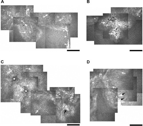
Table 1 Clinical data of four patients with herpes simplex keratitis
Figure 3 In vivo confocal microscopic images at the level of deep epithelium to subbasal nerve plexus in Case 1 (A), Case 2 (B), Case 3 (C), and Case 4 (D).
Notes: Numerous Langerhans cells as dendritic inflammatory cells (arrows) were noted, and were not exclusively located at the dendritic lesion. (Bar =100 μm).
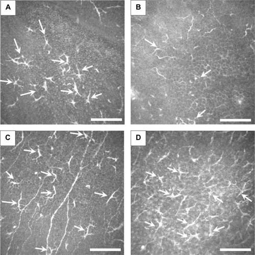
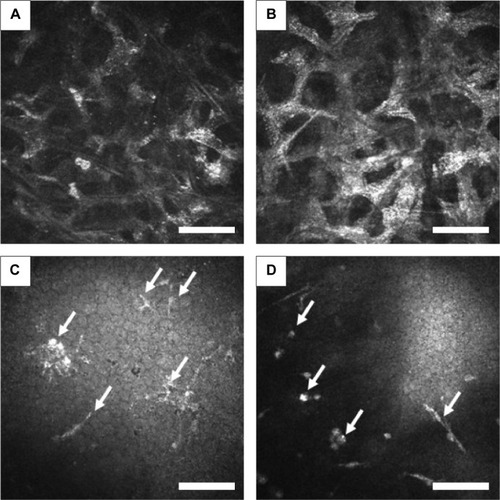
Figure 4 In vivo confocal microscopic images at the level of stroma and endothelium.
Notes: (A and B) Representative stromal image in Case 2 (A) and Case 4 (B). Honeycomb-shaped hyperreflective keratocytes were seen, suggesting stromal edema. (C and D) At the level of endothelium, inflammatory cells (arrows) were seen in Case 1 (C) and 4 (D). (Bar =100 μm).