
Figures & data
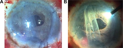
Figure 1 E-DMEK/E-PDEK.
Notes: (A) View of DMEK graft through microscope light shows insufficient visualization; (B) with microscope light turned off and with oblique external illumination by an endoilluminator (vitreo-retinal light pipe), three-dimensional visualization improves with better comprehension of entire graft morphology, orientation, and position.
Abbreviations: E-DMEK, endoilluminator-assisted Descemet membrane endothelial keratoplasty; E-PDEK, endoilluminator-assisted pre-Descemet endothelial keratoplasty.
Abbreviations: E-DMEK, endoilluminator-assisted Descemet membrane endothelial keratoplasty; E-PDEK, endoilluminator-assisted pre-Descemet endothelial keratoplasty.
Video S1 Enhanced visualization and three-dimensional depth perception with the use of oblique external light from the endoilluminator in E-DMEK and E-PDEK.