Figures & data
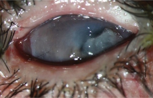
Figure 1 Left eye at presentation (down gaze).
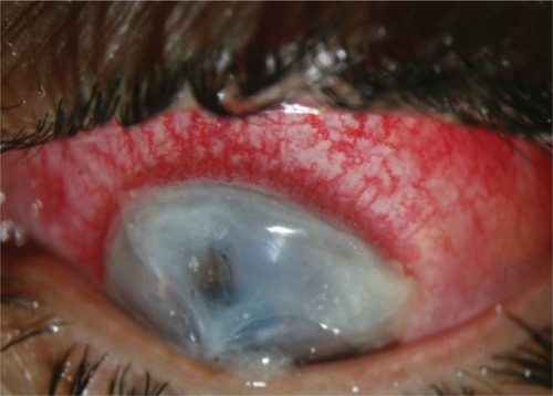
Figure 2 Left eye at presentation (primary position).
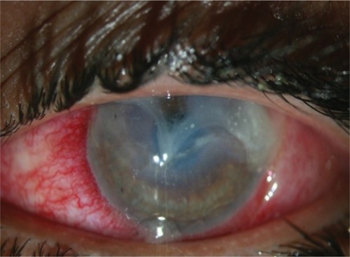
Figure 3 Left eye post penetrating keratoplasty.
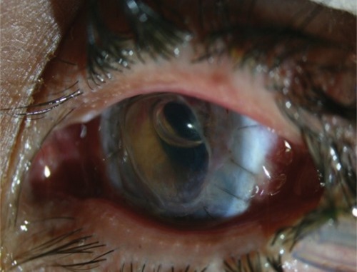
Figure 4 Left eye post lateral tarsorrhaphy.
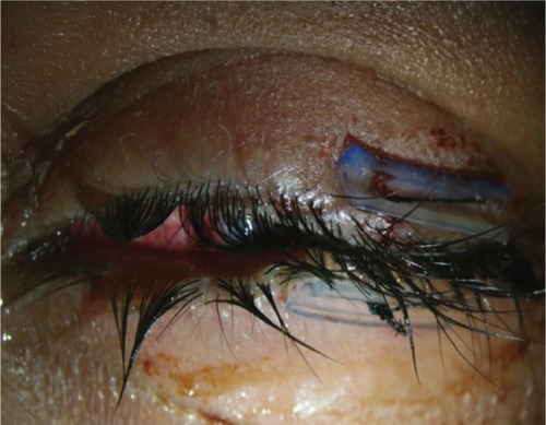
Figure 5 Patient with considerably clear optical zone after penetrating keratoplasty.
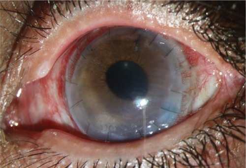
Figure 6 Persistent epithelial defect 3 months after penetrating keratoplasty.
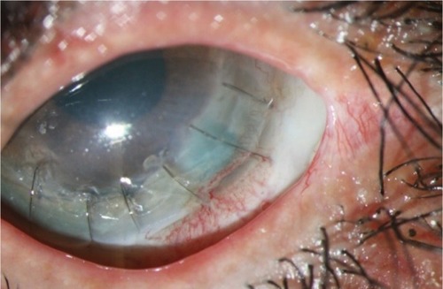
Figure 7 Persistent epithelial defect, fluorescein staining.
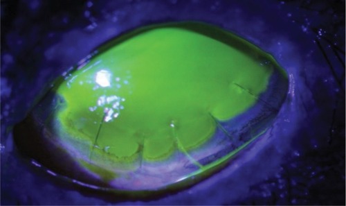
Figure 8 Corneal melting, 6 months later.