Figures & data
Figure 1 Yellow rectangle- Initial selection of both eyeballs on axial view; Red line- Defining the outline of the selected eyeball shape.
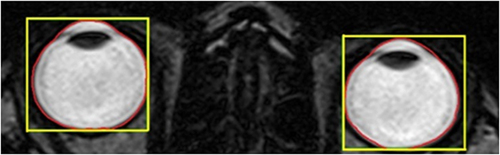
Figure 2 Yellow rectangle- Initial segmentation of the Right eyeball on sagittal view; Red line- Defining the outline of the selected eyeball shape.
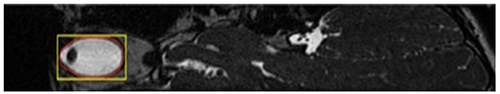
Figure 3 Summary of algorithm for segmentation and measurement of the MRI images.
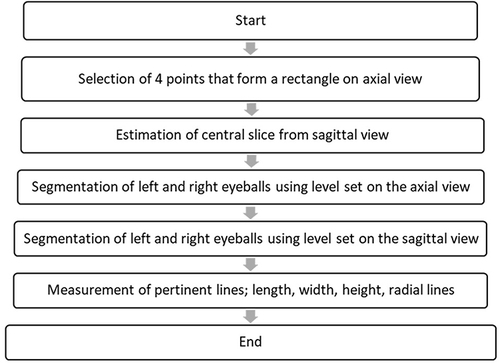
Figure 4 Correlation between LAL measured using ultrasound A-Scan and MRI.
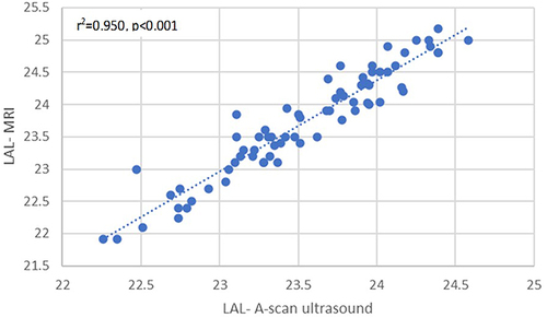
Table 1 Demographic Characteristics of Subjects
Table 2 Summary of Ocular Dimensions in Myopic Children
Table 3 Multivariate Linear Regression Analyses of the Associations of Body Stature Variables with Ocular Dimensions, Corneal Curvature, and SE
Figure 5 Correlation between body height and LAL.
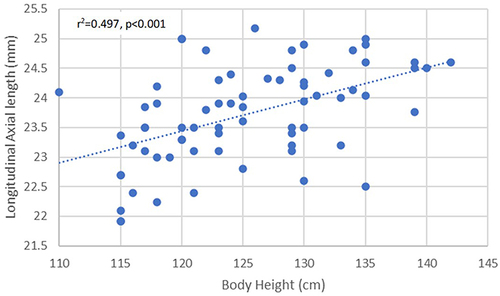
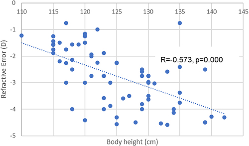
Figure 6 Correlation between body height and refractive error.