Figures & data
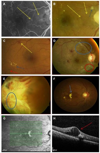
Figure 1 Retinal signs of diabetic retinopathy.
Notes: (A) Fluorescein Angiography (FA) image of Figure 1B, on FA image microaneurysms are readily observed (yellow arrow). (B) Moderate Diabetic retinopathy with exudates in the peri-macula region and no signs of microaneurysms, blue arrow is indicating a cotton wool spot. (C) Moderate diabetic retinopathy with dot (red arrow), blot (yellow arrow) and exudates (blue arrow) present in the posterior pole. (D) Proliferative Diabetic Retinopathy with infarcted neovascularization (red circle) and intraretinal microvascular abnormalities (Blue circle). (E) Tractional Retinal Detachment with proliferative diabetic retinopathy and vitreous hemorrhage, infarcted neo is marked by blue circle. (F) Mild Diabetic retinopathy with clinically significant macula edema, with exudate (blue arrow). (G) Optical coherence image of Figure 1F, showing the exact location of the scan in Figure 1H. (H) The scan line from Figure 1G showing fluid accumulation in the outer plexiform layer of the retina (red arrow), indicating clinically significant macula edema.