Figures & data
Figure 1 (A and B) Location of incision at the level of the pedicle. (C) Triangulation of working and viewing portal above the lamina. (D) Waterflow from the working portal.
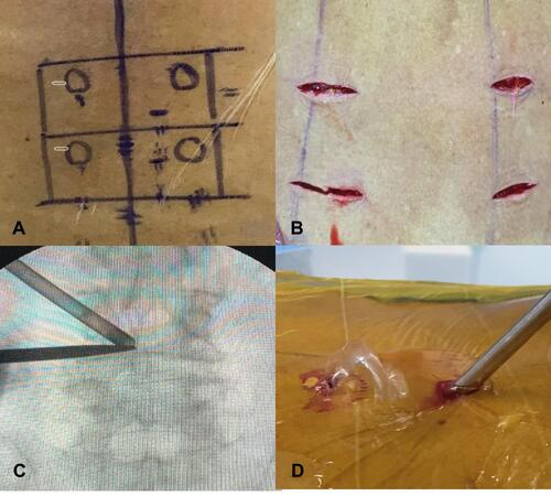
Figure 2 (A) Working position using unilateral biportal endoscopy. (B) Flavectomy piece by piece using Kerrison punch. (C) Contralateral decompression.
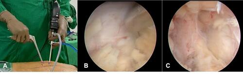
Figure 3 (A) Annulotomy using an annular cutter. (B) Preparation of endplate. (C) Endplate removal completed (asterisk).
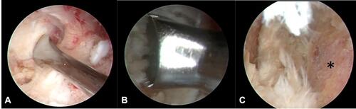
Figure 4 (A) Insertion of reamer under fluoroscopic view. (B) Confirmation on image intensifier.
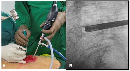
Figure 5 (A) Cage insertion under endoscopic view. (B) Confirmation on image intensifier.
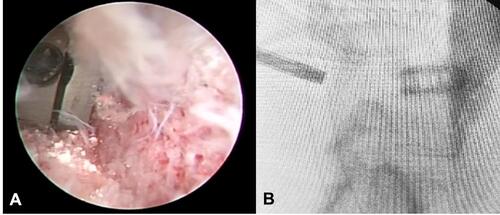
Figure 6 After pedicle screw insertion from AP (A) and lateral (B) view.
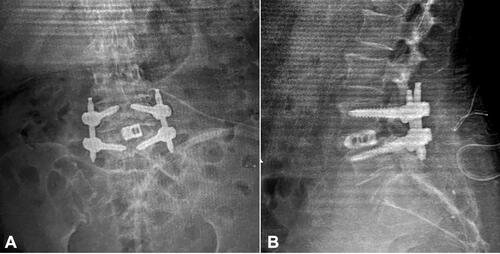
Table 1 General Characteristic of Degenerative Spondylolisthesis Patients Underwent Operations
Table 2 Comparison of Visual Analog Scale Between Conventional MIS TLIF and ULIF
Table 3 Comparison of ODI Between Conventional MIS TLIF and ULIF
Table 4 Comparison of SF-36 Score Between MIS TLIF and ULIF
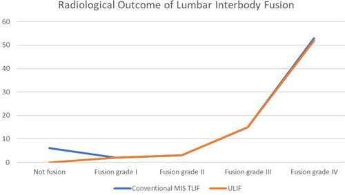
Figure 7 Fusion rate comparison showed no significant difference between ULIF group (orange line) and the conventional MIS-TLIF group (blue line).