
Figures & data
Figure 1 Clusters of round and polygonal vesicular lesions on erythematous to violaceous base at the right hand (A) and post-intravenous acyclovir treatment (B) along with the ulnar nerve distribution with a claw-like deformity of the right hand.
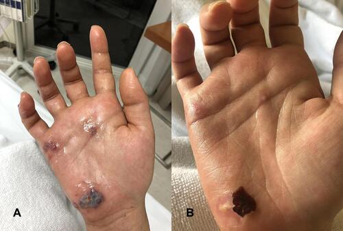
Figure 2 Right ulnar sensory nerve conduction study (NCS) showed no response (yellow highlighting).
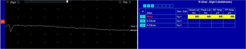
Figure 3 Right 2-channel ulnar motor nerve conduction study (NCS) recorded at the abductor digiti minimi (ADM) and first dorsal interosseous (FDI) muscles, respectively. This demonstrates decreased ADM compound motor action potential (CMAP) amplitude (yellow highlighting) and severely decreased FDI CMAP amplitude (normal ulnar CMAP amplitude in our laboratory is ≥5.0 mV) with a slight decrease in motor nerve conduction velocity (NCV) (yellow highlighting), which can be concluded as axonal involvement of motor fibers, more severe in FDI than ADM muscles.
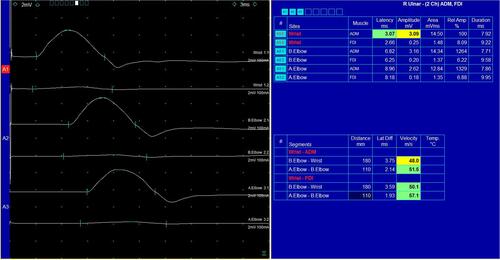
Figure 4 Right median motor nerve conduction study (NCS) recording at abductor pollicis brevis (APB) muscle demonstrated normal compound motor action potential (CMAP) amplitude (normal median CMAP amplitude in our laboratory is ≥5.0 mV) with normal motor nerve conduction velocity (NCV) (green highlighting).
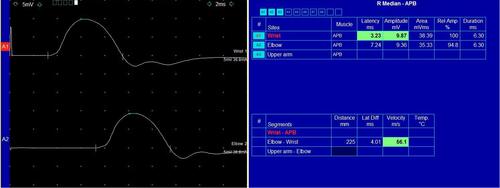
Figure 5 Right claw hand deformity (metacarpophalangeal (MCP) joint hyperextension and interphalangeal (IP) joint slightly flexion) in resting position (A) and after wearing a static figure-of-eight splint for MCP joint flexion (B).
