Figures & data
Figure 1 Measurement of parameters on plain X-rays: (A) shows measurement of the regional kyphotic angle (RA) and vertebral kyphotic angle (VA); (B) shows measurement of percentage of anterior vertebral body height loss (%AVB), which is calculated by the formula: %AVB = 100–2b/(a + c)* 100, where a is the anterior edge height of the vertebra above the fractured vertebra; b is the anterior edge height of the fractured vertebra; and c is the anterior edge height of the vertebra below the fractured vertebra.
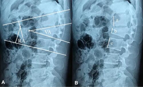
Table 1 Demographic Data
Table 2 Correction Loss of Kyphotic Deformity at Final Follow-Up
Table 3 The Prevalence of Hardware Failure
Table 4 CT Grading of Interbody Fusion Based on Bridwell’s Criteria
Figure 2 Implant failure: (A) breakage of pedicle screws above the fracture level; (B) breakage of pedicle screws below the fracture level; (C) rod pull out of the pedicle screw head.
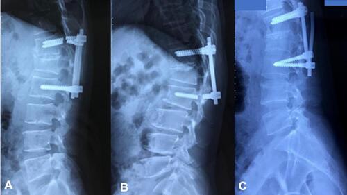
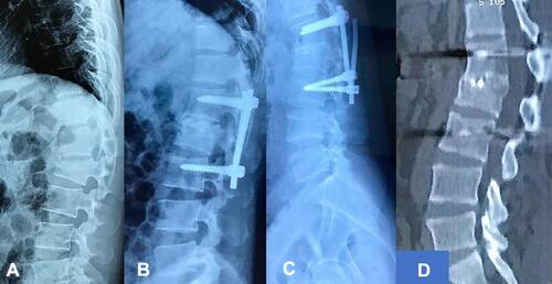
Figure 3 Good bone union in the patient with hardware failure: (A) preoperative X-ray; (B) postoperative X-ray; (C) final follow-up X-ray with hardware failure; (D) sagittal CT at last follow-up with good bone union.