
Figures & data
Figure 1 Histotopograms of peroneal stump ends (haematoxylin and eosin staining. x2.5): (A) Cylindrical shape with formation of bone closure plate from mature bone tissue; (B) Cone-shaped with congestion of the medullary canal (a) and a small periosteal regenerate (b); (C) Cone-shaped with occlusion of the medullary canal (a), violation of the integrity of the cortical diaphyseal layer (arrow); (D) Club-shaped with full length occlusion of the medullary canal with dense fibrous tissue (a), endosteal-periosteal regenerate at the end (b); (E) Club-shaped: resorption of the cortical diaphyseal plate and its replacement by endosteal-periosteal regenerate (a); endosteal bone beams (b); spongy cortical diaphyseal plate in the proximal region (c); fibrous cartilaginous tissue edging the end of the stump (d).
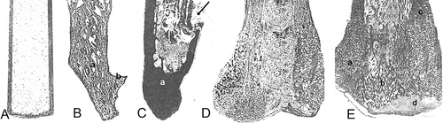
Figure 2 Microphotograph. Dystrophic changes and necrosis of bone tissue. Haematoxylin and eosin staining. x120.
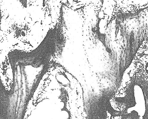
Figure 3 Мicrophotograph. Spongy tissue of periosteal regenerate bordered by hyaline cartilage. Haematoxylin and eosin staining. x90.
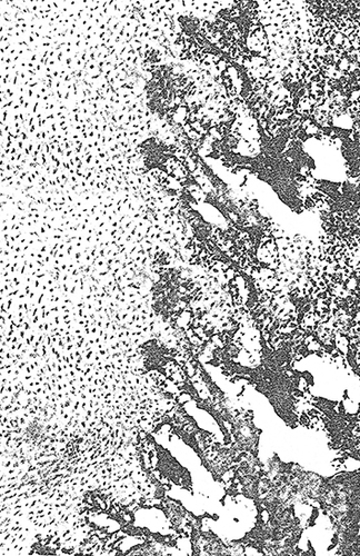
Figure 4 Microphotograph. Dissolution of bone beams of cancellous bone (a) bone-cartilaginous regenerate and its replacement by fibrous tissue (b). Haematoxylin and eosin staining. x130.
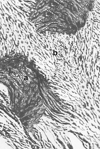
Figure 5 Microphotograph. An area of active osteoclastic resorption of compact bone. Haematoxylin and eosin staining. x120.
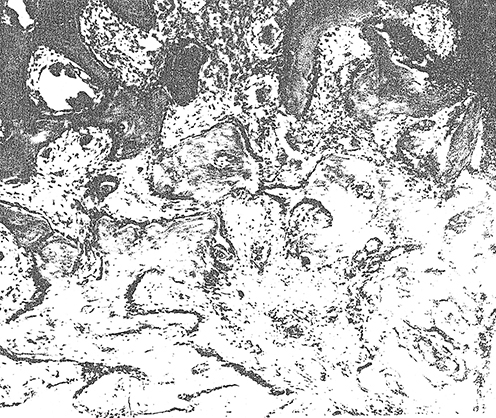
Figure 6 Microphotograph. Resorption of compact bone tissue. Full blood vessels. Haematoxylin and eosin staining. x80.
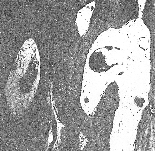
Table 1 Quantitative Evaluation of the Tissue Structures of the Amputation Stump of the Fibula
Table 2 Indicators of the Average Time of Prosthesis Use in Patients of the Study Groups Before and After Surgery