193
Views
2
CrossRef citations to date
0
Altmetric
Review

Rotator cuff repair: challenges and solutions

, , &
Pages 57-69 | Published online: 09 Jul 2015

Figures & data

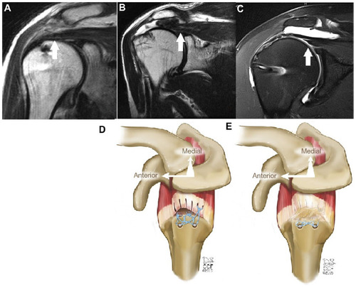
Figure 1 Modes of rotator cuff failure.

Notes: (A) Intact repair on MRI. Note the position of the musculotendinous (MT) junction at the midpoint of the humeral head. (B) Failure with defect on MRI. Note the defect at the greater tuberosity and the significantly retracted position of the MT junction. (C) Failure with continuity on MRI. Note the continuous tissue extending to the greater tuberosity despite significant retraction of the MT junction. White arrows in A–C show position of the MT junction. (D) Illustration of failure with defect. (E) Illustration of failure with continuity. Illustrations by David Schumick, BS, CMI. Reprinted with the permission of the Cleveland Clinic Center for Medical Art and Photography © 2015. All Rights Reserved.
Abbreviation: MRI, magnetic resonance imaging.
Figure 1 Modes of rotator cuff failure.

Table 1 Studies of commercially available scaffolds in augmentation of rotator cuff repairs

Table 2 Studies of commercially available scaffolds in interposition of irreparable rotator cuff tears

Table 3 Level I and II studies on PRP use in rotator cuff repair